
-
Original Article
- Physicochemical and Histomorphometric Comparison of Deproteinized Bovine Bone Graft Materials
- Hyun Wook An, Jin-Young Park, Seon Yeong Kim, Sang Min Lee, Young In Choi, SuJeong Kim, Yuri Kim, Hyun-Chang Lim, Young Woo Song
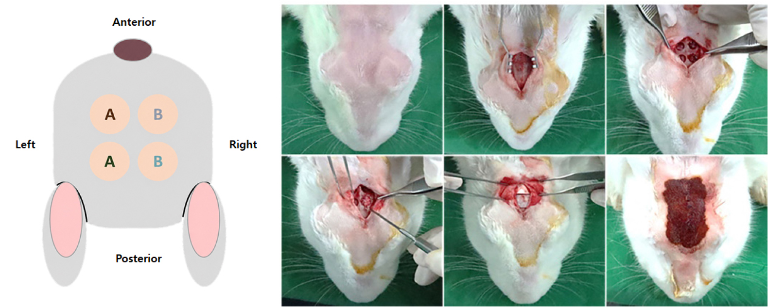
- Purpose: This study compared the physicochemical properties, in vitro biocompatibility, and in vivo bone regeneration of two bovine-derived xenogeneic bone substitutes: Bio-Oss …
- Purpose: This study compared the physicochemical properties, in vitro biocompatibility, and in vivo bone regeneration of two bovine-derived xenogeneic bone substitutes: Bio-Oss and Mega-Oss Bovine Original.Materials and Methods: Physicochemical characterization included SEM, XRD, TEM, FT-IR, thermogravimetric analysis, mercury intrusion porosimetry, and BET analysis. Residual organic components were quantified, and cytotoxicity was assessed using an ISO 10993-5-compliant L-929 MTT assay. In vivo evaluation used a rabbit calvarial defect model (n = 6). Four 6-mm defects per animal were filled with either material and evaluated at 4 and 8 weeks using micro-CT (BV/TV, BMD) and histomorphometric analysis.Results: Both materials exhibited similar physicochemical characteristics, including porous microstructures, low-crystalline hydroxyapatite, and comparable Ca/P ratios and crystallite sizes. Mega-Oss Bovine Original exhibited higher porosity and a greater specific surface area than Bio-Oss. Residual lipid and protein contents were minimal in both materials, and no cytotoxic effects were observed. In the rabbit calvarial defect model, both groups demonstrated favorable tissue compatibility with minimal inflammatory responses. Micro-CT and histomorphometric analyses revealed comparable bone regeneration outcomes between the two groups at both 4 and 8 weeks, with no statistically significant differences in bone volume fraction, bone mineral density, newly formed bone area, or residual graft area (p > .05).Conclusion: Mega-Oss Bovine Original demonstrated physicochemical characteristics, biocompatibility, and bone regeneration performance comparable to those of Bio-Oss. These findings suggest that Mega-Oss Bovine Original may serve as a suitable bovine-derived xenogeneic bone substitute for bone regeneration applications. - COLLAPSE
-
Original Article
- Influence of Scan Body Exposure Height on Digital Impression Accuracy in Maxillary Anterior Implants: A Pilot Study
- Dong Yeob Kang, Seung-Rye Song
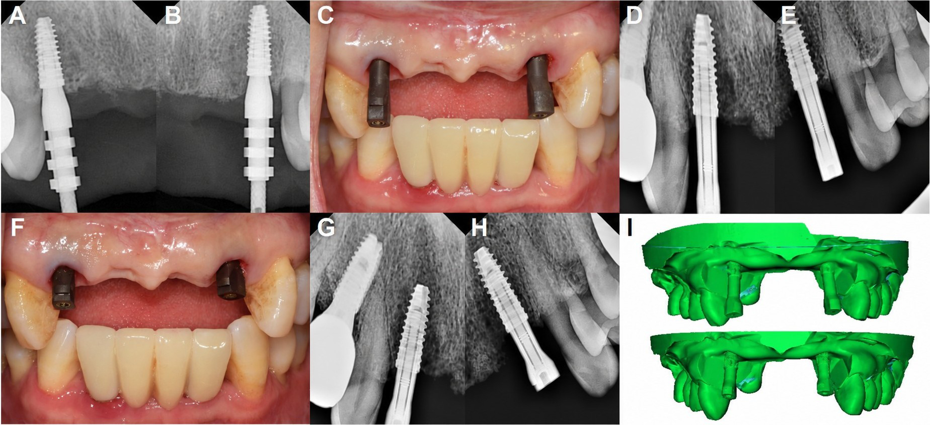
- Purpose: This study evaluated the influence of the supragingival scan body (SB) exposure height on the accuracy of intraoral digital impressions at …
- Purpose: This study evaluated the influence of the supragingival scan body (SB) exposure height on the accuracy of intraoral digital impressions at maxillary anterior implant sites.Materials and Methods: Digital impression datasets from two maxillary anterior implant sites were retrospectively analyzed. A Short SB scan was performed during the provisional restoration stage, whereas a Long SB scan was performed during the definitive restoration stage. A stone cast was fabricated from a provisional stage open-tray polyvinyl siloxane impression, and the same cast was digitized with each physical SB seated to generate condition-specific reference datasets. The iterative closest point (ICP) registration root mean square (RMS) error was recorded, and the linear deviation (LD) and angular deviation (AD) were obtained using Random Sample Consensus (RANSAC)-based cylinder fitting. Statistical analyses were not performed due to the single-patient design of the study.Results: The mean supragingival SB exposure heights were 6.39 mm and 2.73 mm for the Long SB and Short SB conditions, respectively. The Long SB condition demonstrated lower ICP registration RMS error (29.8 µm) than the Short SB condition (70.4 µm). Mean LD values were 1.17 and 1.35 mm, and mean AD values were 0.85° and 3.68° for the Long SB and Short SB conditions, respectively.Conclusion: In this retrospective single-patient pilot study, the dataset with reduced supragingival SB exposure height showed greater AD and higher ICP registration RMS errors at the maxillary anterior implant sites. Although no causal relationship can be established from these findings, the supragingival SB exposure height may warrant further investigation in relation to digital implant impression accuracy. - COLLAPSE
-
Clinical or Case Report
- Management of Narrow Alveolar Ridges Using a Modified Multistaged Alveolar Ridge-Splitting Technique with Miniplates and Screws: A Case Series
- Heuiyung Oh, Minwoo Oh
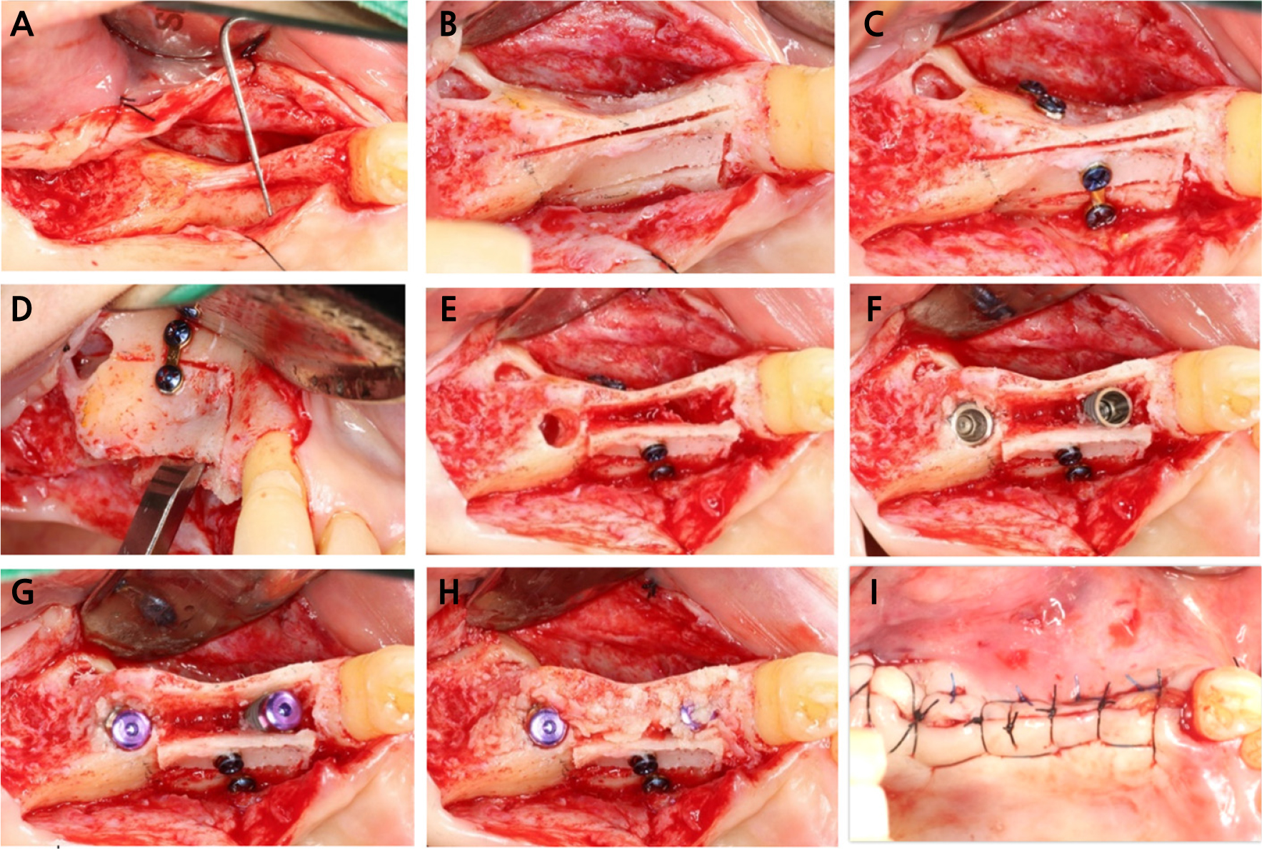
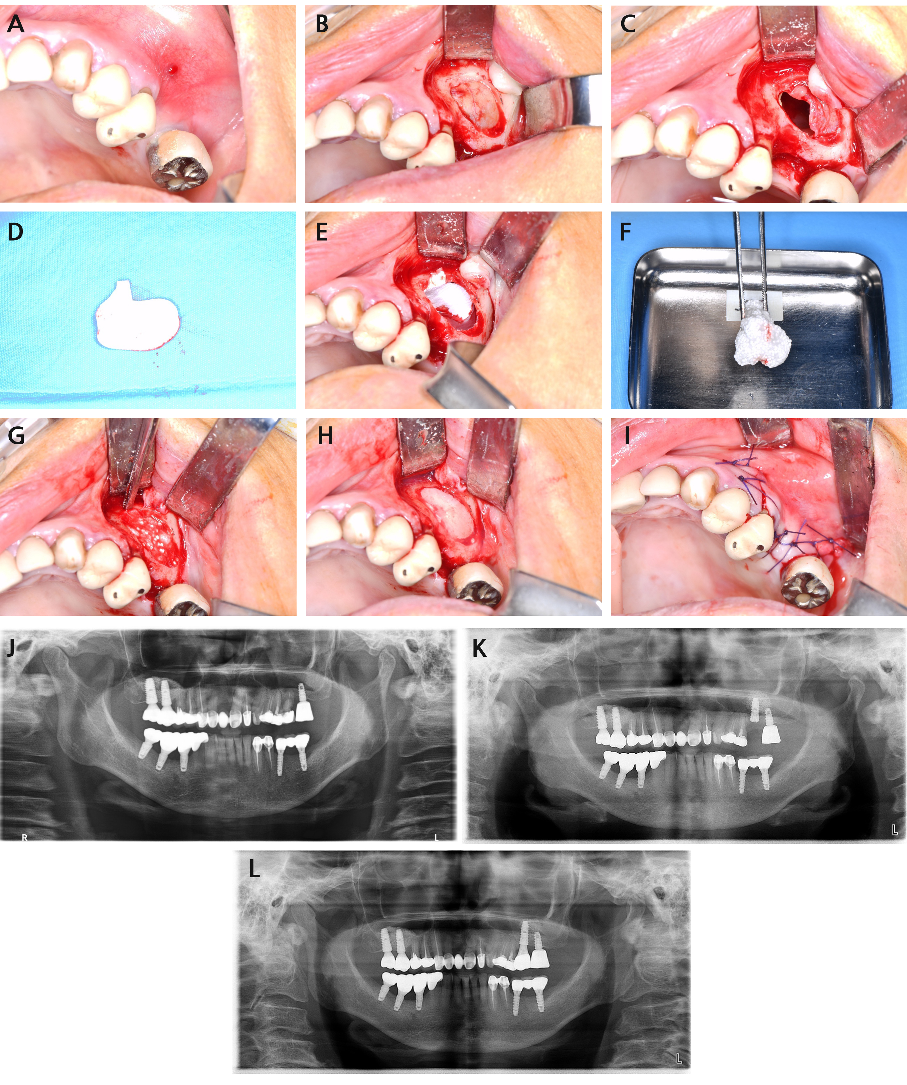
- Clinicians commonly face horizontal alveolar ridge deficiency when planning for dental implant placement. Alveolar ridge splitting is a highly predictable surgical technique …
- Clinicians commonly face horizontal alveolar ridge deficiency when planning for dental implant placement. Alveolar ridge splitting is a highly predictable surgical technique used to increase horizontal alveolar bone width. This report describes three clinical cases in which a modified alveolar ridge-splitting technique was performed using miniplates and screws. Clinical and radiographic evaluations demonstrated post-extraction horizontal ridge deficiency in all three patients. A modified, staged ridge-splitting technique using miniplates and screws was employed to facilitate implant placement with adequate bone width. Controlled ridge expansion and primary implant stability were achieved in all cases, and healing proceeded without complications. Subsequent clinical and radiographic assessments confirmed stable marginal bone levels and successful osseointegration. Within the limitations of this case series, modified alveolar ridge splitting using miniplates and screws proved to be an effective approach for horizontal ridge augmentation, enabling simultaneous implant placement in narrow ridges. - COLLAPSE
-
Clinical or Case Report
- Staged Interdisciplinary Treatment for Maxillary Central Incisor Implant Rehabilitation with Crowding and Severe Alveolar Defect: A Clinical Report
- Minhee An, Sunjai Kim, Jae-Seung Chang, Se-Wook Pyo
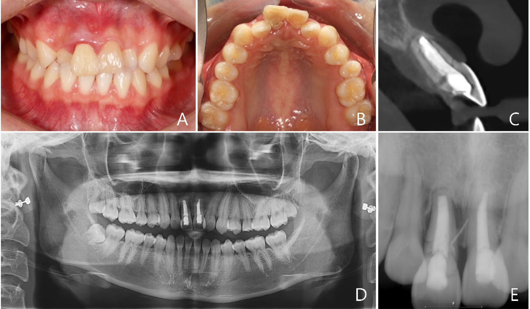
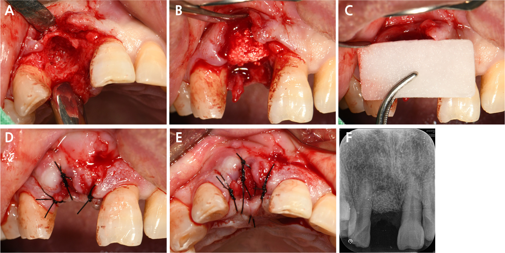
- Implant rehabilitation of a maxillary central incisor in the esthetic zone is often challenging when severe alveolar bone loss is accompanied by …
- Implant rehabilitation of a maxillary central incisor in the esthetic zone is often challenging when severe alveolar bone loss is accompanied by inadequate restorative space owing to crowding. This clinical report describes the staged interdisciplinary treatment of a 20-year-old female presenting a crown-to-root fracture of the maxillary right central incisor, severe alveolar bone defect, and limited restorative space caused by anterior crowding. Following tooth extraction, alveolar ridge preservation (ARP) was performed to minimize ridge collapse, and orthodontic treatment was initiated to establish adequate space and alignment for prosthetically driven implant placement. Despite partial ridge preservation, additional site development with block bone grafting was required because of insufficient bone volume for implantation. After adequate bone regeneration, implant placement and prosthetic rehabilitation were completed with favorable esthetic and functional outcomes. This case demonstrates that a staged interdisciplinary treatment involving space creation and site development may facilitate implant rehabilitation in compromised anterior maxillary conditions. - COLLAPSE
-
Clinical or Case Report
- Clinical Application of a Porcine-Derived Collagen Matrix: A Case Series
- Woo-Taek Song, Ji-Hun Park, Hyun-Jung Oh, Bo-Ah Lee, Young-Taek Kim
- Restoration of soft tissue defects is essential for periodontal and implant surgeries. Although autologous grafts remain the gold standard, they increase patient …
- Restoration of soft tissue defects is essential for periodontal and implant surgeries. Although autologous grafts remain the gold standard, they increase patient morbidity owing to donor site harvesting. This retrospective case series evaluated the porcine-derived collagen matrix as a less invasive alternative across three indications: vestibuloplasty, soft tissue augmentation, and alveolar ridge preservation (ARP). The xenogeneic matrix showed biocompatibility, favorable initial healing, and rapid epithelialization. The elimination of donor site surgery reduced patient morbidity. The matrix was integrated without notable complications, providing acceptable esthetic and functional contours for both open and closed healing. However, physical limitations including postoperative absorption, decreased tensile strength upon hydration, and rapid degradation were observed as a biomechanical trade-off. In conclusion, this matrix serves as a clinically feasible substitute for autogenous grafts, minimizing discomfort while achieving favorable outcomes. - COLLAPSE
-
Dental Technique
- Resorbable Collagen Membrane and Fibrin Glue Application with a Handle Design for Repairing Sinus Membrane Perforation
- Ikhyeon Kim, Seunghun Baek, Kang-Min Ahn
- Perforation of the Schneiderian membrane during lateral sinus floor elevation is a frequent intraoperative complication that can compromise graft stability and peri-implant …
- Perforation of the Schneiderian membrane during lateral sinus floor elevation is a frequent intraoperative complication that can compromise graft stability and peri-implant health. This technical note describes a modified membrane application technique utilizing an ergonomic “handle” design and fibrin glue for perforation repair and demonstrates its clinical application. A 71-year-old woman with a history of osteoporosis and denosumab therapy experienced membrane perforation during lateral sinus augmentation and simultaneous implant placement at site #26. During the procedure, the sinus membrane was further elevated to reduce the functional defect size. A resorbable collagen membrane was custom-trimmed to include a protruding “handle”, enabling precise, guided insertion over the defect using surgical forceps to prevent inward displacement. A cohesive mass of the synthetic bone graft material, rhBMP-2, and fibrin glue was safely packed into the subantral space, followed by tack-free repositioning and initial stabilization of the lateral bony window using additional fibrin glue. This technique successfully secured the repair site and prevented particle scattering. Compared with conventional approaches, this modified approach may provide superior mechanical stability and simplifies the overall procedure. However, long-term follow-up and histological evaluation are required to confirm the stability and quality of the newly formed bone at the perforated site. - COLLAPSE

Journal Informaiton
 Journal of implantology and applied sciences
Journal of implantology and applied sciences


대한구강악안면임플란트학회
(Yeoksam-Dong,KFOST) New-building room507, 22, Teheran-ro 7-gil, Gangnam-gu, Seoul, 06130, Korea
Tel: +82-2-558-5966 / Fax: +82-2-558-5965 / E-mail: webmaster@implant.or.kr Copyright© The Korean Academy of Oral & Maxillofacial Implantology. Powered by APUB
Tel: +82-2-558-5966 / Fax: +82-2-558-5965 / E-mail: webmaster@implant.or.kr Copyright© The Korean Academy of Oral & Maxillofacial Implantology. Powered by APUB