


Ⅰ. Introduction
Ⅱ. Materials and Methods
1. Sample Preparation
2. Physicochemical Characterization
3. In Vitro Cytotoxicity Evaluation
4. In Vivo Study
Ⅲ. Results
1. Material Characterization
2. In vitro cytotoxicity evaluation
3. In Vivo Evaluation
Ⅳ. Discussion
Ⅴ. Conclusion
Ⅰ. Introduction
Bone grafting has become an essential therapeutic approach in dentistry for reconstructing bone defects caused by periodontal disease and trauma, with more than 2 million procedures performed annually worldwide.1
Among the three key components of tissue engineering—scaffolds, cells, and growth factors—bone graft materials play a fundamental role by offering a three-dimensional structural framework that supports new bone formation and enhances tissue regeneration.
Among bone graft materials, autogenous bone has long been considered the gold standard because of its superior osteogenic and osteoinductive properties. However, it has well-recognized limitations, including the need for an additional surgical procedure to harvest donor tissue and the limited availability of graft material. To overcome these drawbacks, synthetic bone substitutes have been developed and demonstrated clinically meaningful outcomes; however, their function is generally limited to osteoconductive support.2,3,4
In contrast, deproteinized bovine bone mineral (DBBM), a xenogeneic graft material, has a calcium-to-phosphorus (Ca/P) ratio comparable to human bone. Through a specialized processing method that removes organic components, DBBM retains the natural porous architecture of native bone tissue, representing a significant advantage.5,6,7 In particular, Bio-Oss (Geistlich Pharma AG, Wolhusen, Switzerland), widely recognized as a global benchmark, has demonstrated excellent biocompatibility and osteoconductive properties through decades of clinical evidence.
Therefore, direct comparative studies with established reference products are essential to validate the clinical efficacy of newly developed bone graft materials. This study aims to examine the physicochemical characteristics and in vivo bone regeneration performance, via histomorphometric analysis in a rabbit calvarial defect model, of a novel bovine-derived xenograft produced using a proprietary manufacturing process, compared with Bio-Oss. This study seeks to determine whether the newly developed xenograft demonstrates equivalent or superior performance to the reference product and provides scientific evidence supporting its potential clinical use.
Ⅱ. Materials and Methods
1. Sample Preparation
In this study, Mega-Oss Bovine Original (MEGAGEN Implant Co., Ltd., Daegu, South Korea) served as the experimental group, while Bio-Oss was selected as the control group.
Mega-Oss Bovine Original was manufactured using a proprietary physicochemical process intended to facilitate the removal of bacteria, viruses, and infectious proteins. Residual organic components were further reduced through low-temperature heat treatment. The material was subsequently sterilized using gamma irradiation after packaging.
Bio-Oss is a natural inorganic bone graft material derived from bovine bone with its organic components removed. It has a porous structure similar to human bone. The commercially available product was used as provided.
2. Physicochemical Characterization
1) Morphological Analysis
Surface morphology and microstructural characteristics were examined using scanning electron microscopy (AIS1800C; Seron Technologies Inc., Uiwang, South Korea). Images were captured at magnifications of ×100, ×500, and ×3,000, depending on the observation purpose.
2) Crystallographic Analysis
(1) Crystallinity and Ca/P ratio were analyzed using X-ray diffraction (XRD; Miniflex600, Rigaku, Japan), and crystallite size was semiquantitatively estimated using the Scherrer equation:
where λ is the X-ray wavelength, β is the full width at half maximum, and θ is the Bragg angle.
(2) Transmission electron microscopy (Tecnai G2 F20 S-Twin; FEI Company, Hillsboro, USA) was performed at an accelerating voltage of 200 kV to observe the crystal morphology and sizes.
3) Functional Group and Chemical Structure Analysis
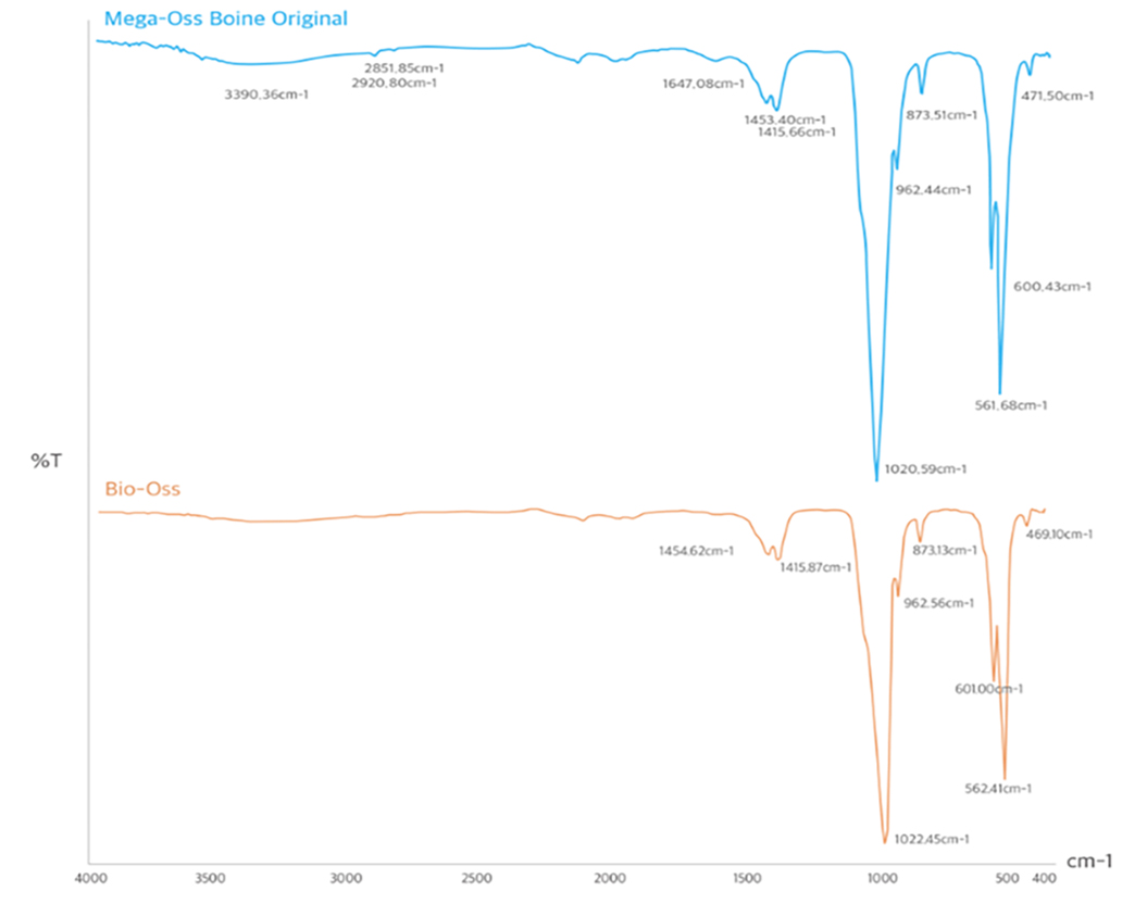
FT-IR (Frontier; PerkinElmer, Waltham, USA) was performed over the mid-infrared range of 400–4000 cm‒1 to identify organic components and characteristic hydroxyapatite(HA) absorption bands.
4) Porosity and Specific Surface Area Analysis
Mercury intrusion porosimetry (AutoPore V 9620; Micromeritics Instrument Corp., Norcross, USA) was used to measure porosity based on the assumption that cylindrical pores exist within the sample and that liquid penetration into smaller pores follows capillary principles.8 For nonwetting fluids, such as mercury, the pore diameter is determined based on the following relationship:
where D is the pore diameter (nm), P is the applied pressure (psi), γ is the surface tension of the fluid (mJ·m‒2), and θ is the contact angle between the solid and fluid.
Specific surface area was measured using nitrogen adsorption and calculated from the adsorption isotherm with the Brunauer–Emmett–Teller method using a surface area and porosity analyzer (Quadrasorb evo; Quantachrome Instruments, Boynton Beach, USA).
5) Analysis of Organic Content
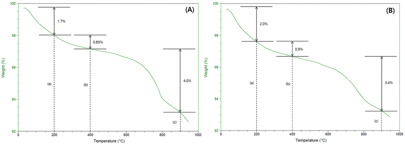
(1) Thermogravimetric analysis (Pyris Diamond TGA; PerkinElmer) was performed to assess weight changes with a temperature range of 25–950°C at a constant heating rate of 10°C /min.
(2) A Soxhlet extraction system (Soxtherm 416; C. Gerhardt GmbH & Co. KG, Königswinter, Germany) was used to quantify residual lipid content. Protein content was determined using the Kjeldahl method with a nitrogen/protein analyzer (Kjeltec 8400; FOSS Analytical A/S, Hillerød, Denmark), a widely recognized standard method for nitrogen analysis. The Kjeldahl method allows conversion of protein nitrogen into ammonium without interference from residual lipid components or other constituents found in the sample.9,10,11
3. In Vitro Cytotoxicity Evaluation
Cytotoxicity was assessed based on ISO 10993-5 using mouse fibroblast L-929 cells, derived from the connective tissue of a 100-day-old male mouse.
Cells were cultured in RPMI medium (Gibco; Life Technologies, Grand Island, USA) supplemented with 10% fetal bovine serum (Gibco; Life Technologies) in a CO2 incubator at 37°C with 5% CO2 (MCO-15AC; SANYO, Osaka, Japan). Sample extracts were prepared by incubating the materials in RPMI medium with 10% FBS at 37°C for 72 h under continuous agitation.
The fibroblast suspension was adjusted to 2.0 × 10⁵ cells/mL, and 0.2 mL was seeded into each well of a 48-well plate, followed by 24h incubation. The culture medium was then removed and replaced with 0.2 mL of the sample extract per well, followed by an additional 24 h incubation.
After incubation, the extracts were removed, and 0.2 mL of RPMI solution containing penicillin and MTT reagent (3-(4,5-dimethylthiazol-2-yl)-2,5-diphenyl tetrazolium bromide) was added to each well followed by 3h incubation. The MTT solution was subsequently discarded, and 0.2 mL of dimethyl sulfoxide was added to dissolve the formazan crystals. Absorbance was measured at 570 and 650 nm using a microplate spectrophotometer (PowerWave XS; BioTek Instruments, Winooski, USA), and cell viability was calculated relative to the control group.
4. In Vivo Study
1) Surgical Procedure
Bone regeneration was evaluated using a rabbit calvarial defect model, with all surgical and animal care procedures conducted under an approved Institutional Animal Care and Use Committee protocol (IACUC; Approval No.: KMEDI-22042705-00). The outcomes of the present experiment were addressed according to the ARRIVE (Animal Research: Reporting of In Vivo Experiment) guidelines. This study used six mature male New Zealand white rabbits (mean body weight: 2.6 kg) (Table 1).
Table 1.
Experimental groups
| Group | Animal ID | Time point |
| G1 | #1101-#1103 | 4 weeks |
| G2 | #1201-#1203 | 8 weeks |
Preoperatively, the animals were acclimated under controlled environmental conditions with a standardized diet and housing.
General anesthesia was induced by intramuscular injection of ketamine (35 mg/kg) and xylazine (Rompun, 5 mg/kg). Meloxicam (0.2 mg/kg) and enrofloxacin (10 mg/kg) were administered subcutaneously to prevent postoperative inflammation and infection.
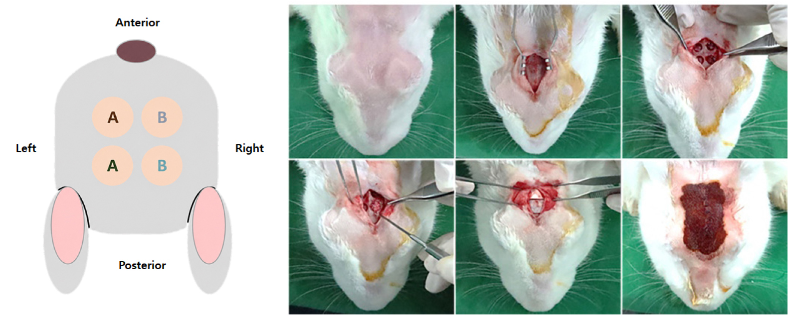
After shaving and disinfecting the surgical site with povidone-iodine and alcohol, a skin incision was made, and the periosteum was elevated to expose the calvarial bone. Four 6 mm diameter circular defects were created per animal using a surgical motor and trephine bur, with reference to the sagittal suture to ensure standardized positioning.
Two defects were filled with Bio-Oss (particle size: 0.25–1.0 mm), and the remaining two with Mega-Oss Bovine Original (particle size: 0.25–1.0 mm). Following graft placement, a resorbable collagen membrane (Genoss Co., Ltd., Suwon, South Korea) was adapted to the defect and positioned to completely cover the grafted area. The periosteum was subsequently sutured to stabilize the graft and membrane, followed by closure of the skin. The surgical site was disinfected with povidone-iodine (Fig. 1).
During 4 and 8 weeks postimplantation, animals were euthanized under anesthesia by rapid intravenous potassium chloride injection. Death was confirmed by cessation of respiration and heartbeat before specimen harvesting.
2) In Vivo Evaluation
(1) Gross Evaluation
Animals were observed once daily for general clinical signs and mortality and any abnormal findings, including the type of symptom, onset time, and severity, were recorded.
(2) Radiographic Evaluation
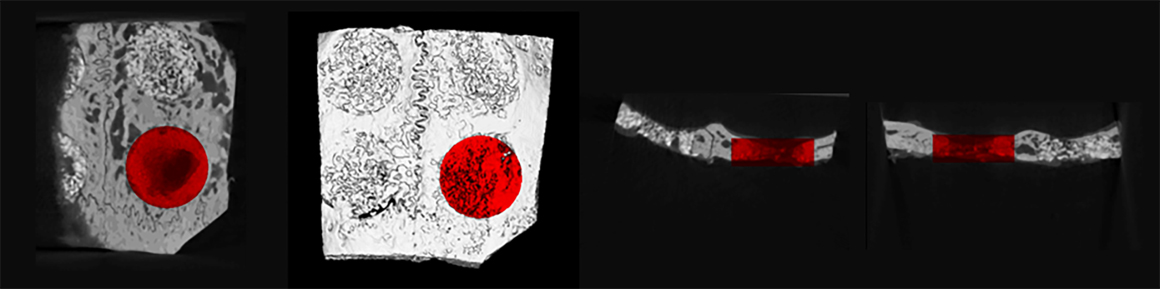
At 4 and 8 weeks, calvarial specimens were harvested and scanned using microcomputed tomography (Quantum FX; PerkinElmer). Image analysis was performed using Analyze 12.0 software by applying a region of interest with a 5.625 mm diameter and a 1.875 mm height. Bone regeneration was quantitatively evaluated by measuring bone volume/total tissue volume (BV/TV, %) and bone mineral density (BMD, mg/cc) (Fig. 2).
(3) Histological and Histomorphometric Evaluation
After micro-CT analysis, specimens were fixed in 10% formalin and decalcified in 10% EDTA solution at 37°C for 4 weeks. The tissues were trimmed, paraffin-embedded and sectioned for slide preparation.
Sections were stained with hematoxylin and eosin (H&E) and Masson’s trichrome. Histopathological evaluation was performed on H&E-stained sections to examine new bone formation and inflammatory response around the graft materials.
Tissue responses at the implantation site were assessed using a modified scoring system based on ISO 10993-6 guidelines for bone graft materials (Table 2). Neovascularization and fibrosis were assessed as tissue response endpoints; however, these findings were interpreted as part of the normal healing and regenerative processes of the cranial defect rather than graft-induced irritation. Histomorphometric analysis was performed using the i-Solution Lite image analysis system (IMT i-Solution Inc., Vancouver, Canada). The region of interest (ROI) was manually delineated by tracing the margins of the original surgically created cranial defect and encompassed the entire defect area bounded by the native calvarial bone. Newly formed bone was identified as mineralized tissue containing osteocytes within lacunae and exhibiting morphological characteristics of bone. Bone regeneration was expressed as the total area of newly formed bone and as a percentage of the total defect area. Residual graft material was quantified within the same ROI and identified based on its characteristic polygonal morphology and staining properties. Areas representing only the outlines of previously present graft particles without remaining material were excluded from analysis. All measurements were performed by a single blinded examiner using predefined criteria.
Table 2.
Semi-quantitative evaluation system for biological effects after implantation based on ISO 10993-6
(4) Statistical analysis
Because four defects were created within each rabbit, the animal was considered the experimental unit for all in vivo analyses. Measurements obtained from individual defects were averaged for each animal prior to statistical analysis to account for within-animal clustering and avoid pseudoreplication. Two-way analysis of variance (ANOVA) was used to evaluate the effects of treatment group and healing period on micro-CT parameters (BV/TV and BMD) and histomorphometric outcomes (new bone area and residual graft area). Data are presented as mean ± standard deviation (SD). Statistical significance was defined as p < .05.
Ⅲ. Results
1. Material Characterization
1) SEM
Microstructural observation using SEM showed that Mega-Oss Bovine Original (Figs. 3A to 3C) and Bio-Oss (Figs. 3D to 3F) had highly similar surface morphologies and pore structures across all magnifications.
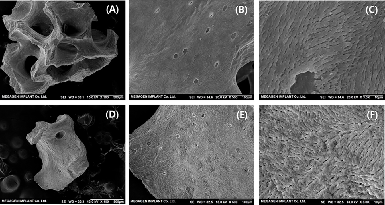
At low magnification (×100), both materials exhibited well-developed macropores (>100 μm), conducive to new bone formation and vascular infiltration (Figs. 3A and 3D).
2) XRD (X-ray Diffraction)
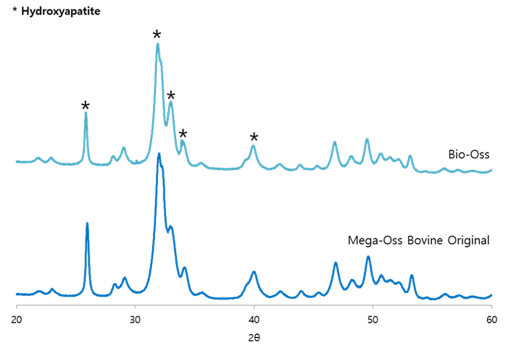
XRD analysis showed that both Mega-Oss Bovine Original and Bio-Oss exhibited diffraction peaks corresponding to the standard HA pattern, confirming an identical crystalline phase (Fig. 4).
Notably, both samples revealed broad diffraction peaks, characteristic of low-crystalline HA.12
The Ca/P ratio of Mega-Oss Bovine Original was 1.66, comparable to Bio-Oss (1.67) and close to the theoretical value of human bone.13 In addition, the crystallite size calculated using the Scherrer equation was 9.79 nm for Mega-Oss Bovine Original, comparable to that of Bio-Oss (10.79 nm).
3) TEM
High-resolution TEM images revealed that Mega-Oss Bovine Original (Figs. 5A and 5B) and Bio-Oss (Figs. 5C and 5D) are composed of densely aggregated nanocrystals.
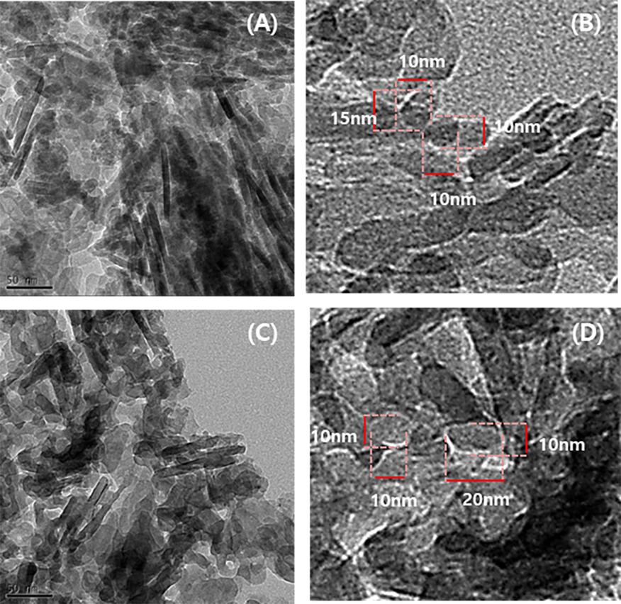
Morphological analysis revealed a mixture of plate-like, spherical (or hemispherical), and rod- shaped crystal structures in both materials. These features are consistent with the typical nanostructure of low-crystalline HA found in natural bone tissue.14
The crystallite size distribution ranged from approximately 10 to 30 nm in both samples. This is comparable to the reported values for human bone (length: 21 ± 7 nm, width: 6 ± 1 nm) and bovine bone (length: 13 ± 2 nm, width: 7 ± 1 nm).15
Notably, Mega-Oss Bovine Original exhibited particle morphology and crystallite size distributions similar to those observed in Bio-Oss, suggesting comparable nanoscale biomimetic characteristics.
4) FT-IR
FT-IR analysis was conducted to identify inorganic composition and detect the presence of residual organic components.16
Both Mega-Oss Bovine Original and Bio-Oss exhibited matching fingerprint regions corresponding to HA (Fig. 6). Characteristic phosphate (PO43‒) stretching bands were observed at approximately 962 and 1020 cm‒1, while bending bands appeared at 561–562 cm‒1 and 600–601 cm‒1.
In addition, carbonate (CO32‒) absorption bands at 873, 1414–1415, and 1453–1454 cm‒1 were observed in both materials, indicating carbonated HA similar to that of natural bone.
Importantly, amide I and II bands (approximately 1550–1650 cm‒1), which are indicative of protein content, were not detected in either sample, suggesting substantial removal of organic components.
5) Porosity and Specific Surface Area
While SEM provided qualitative insights into microstructure, quantitative analysis of porosity was performed using mercury intrusion porosimetry and the BET method.
Mega-Oss Bovine Original exhibited a porosity of 86.65%, compared with 71.61% for Bio-Oss. Furthermore, the specific surface area of Mega-Oss Bovine Original was measured at 127.015 m2/g, approximately 38% higher than that of Bio-Oss (91.965 m2/g).
The average pore diameter was 15.20 nm, revealing a well-developed nanoporous structure (Table 3).
Table 3.
Porosity and specific surface area of the samples
| Product | Porosity | Surface area | Average pore diameter |
| Bio-Oss | 71.61% | 91.965 m2/g | 19.78 nm |
| Mega-Oss Bovine Original | 86.65% | 127.015 m2/g | 15.20 nm |
6) TGA
TGA results showed that Mega-Oss Bovine Original and Bio-Oss exhibited nearly identical thermal decomposition profiles over the entire temperature range from room temperature to 900°C (Fig. 7).
Both samples demonstrated a high residual mass of over 93%, indicating excellent thermal stability and preservation of physicochemical structure.
An initial weight loss of approximately 1.7–2.0% up to 200°C was attributed to the removal of adsorbed moisture. Notably, in the temperature range of 200–400°C, which corresponds to the primary decomposition region of organic components and proteins, both materials showed minimal weight loss (<1%; 0.85–0.9%).
Further gradual weight loss (3.4–4.0%) up to 900°C is considered to result from the decomposition of carbonate (CO32⁻) groups and structural transitions within HA.
7) Organic Content
Complete removal of organic components during the manufacturing process is critical to prevent potential inflammatory and immunological responses after implantation.
In this study, the residual organic content of Mega-Oss Bovine Original was analyzed to verify the effectiveness of the proprietary purification process. Mega-Oss Bovine Original exhibited lipid and protein contents of 0.06% and 0.03%, respectively, Bio-Oss exhibited corresponding values of 0.07% and 0.04%.
2. In vitro cytotoxicity evaluation
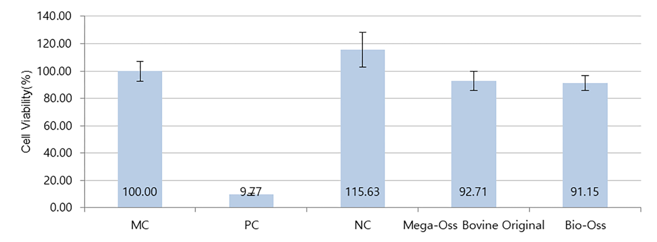
Cytocompatibility of the bone graft materials was evaluated using an MTT extract assay with L-929 fibroblasts, and the results are presented in Fig. 8.
The positive control (PC) and negative control (NC) showed cell viabilities of 9.77% and 115.63%, respectively, demonstrating that the assay met the acceptance criteria and was therefore considered valid. The cell viabilities of the Mega-Oss Bovine Original and Bio-Oss groups were 92.71% and 91.15%, respectively, indicating that both materials exhibited high and comparable cell viability. Furthermore, both materials demonstrated cell viability well above the 70% cytotoxicity threshold defined in ISO 10993-5, confirming their noncytotoxic nature.
3. In Vivo Evaluation
A 6 mm rabbit calvarial defect model was selected because it has been widely used in preclinical studies for the evaluation of bone graft materials and bone regeneration. Although this defect size is frequently regarded as a critical-size defect, its classification may vary depending on the experimental conditions and healing period.17,18,19 In addition, the absence of an untreated control defect limits the ability to distinguish bone formation induced by the graft materials from spontaneous healing. Therefore, the results should be interpreted with caution
1) Gross Evaluation
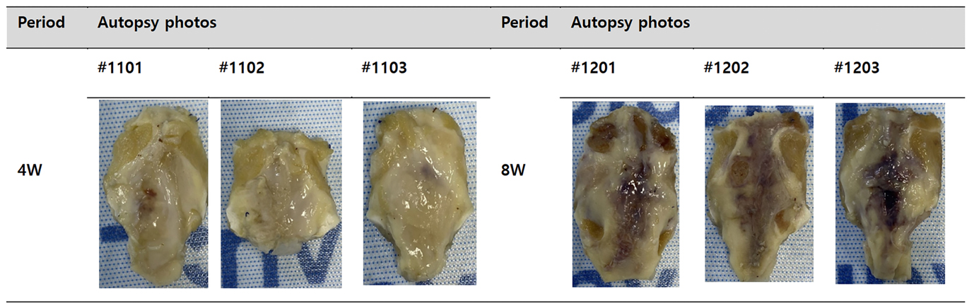
No signs of severe inflammation, infection, or tissue necrosis were observed at the implantation sites in either the Mega-Oss Bovine Original or Bio-Oss groups (Fig. 9).
2) Micro-CT Analysis
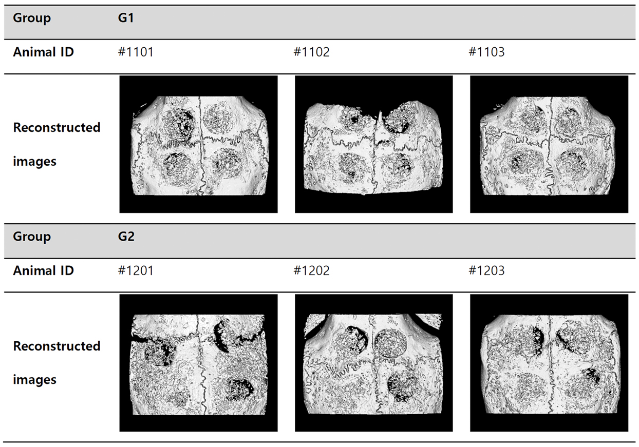
Micro-CT analysis was performed at 4 and 8 weeks post-implantation in separate experimental groups to evaluate mineralized tissue formation and residual graft presence within the defect sites. At both time points, mineralized tissue formation and residual graft materials were observed in all samples. At 4 weeks, the defect margins remained clearly distinguishable, and no marked intergroup differences were identified. At 8 weeks, mineralized tissue occupied a larger portion of the defect area compared with the 4-week time point in both groups, and the overall pattern of mineralized tissue formation appeared similar between the Mega-Oss Bovine Original and Bio-Oss groups (Fig. 10).
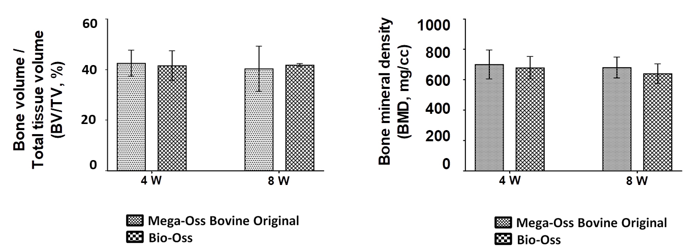
Quantitative analysis of BV/TV and BMD demonstrated that the Bio-Oss and Mega-Oss Bovine Original groups exhibited comparable BV/TV and BMD values at both 4 and 8 weeks. No statistically significant differences were observed between the two groups for either parameter (p > .05). Due to specimen damage during preparation, some samples from animal #1102 were excluded from the micro-CT analysis, and quantitative measurements were therefore performed using the remaining specimens. Specifically, the BV/TV values were 41.59 ± 5.91% and 42.56 ± 5.12% at 4 weeks and 41.80 ± 0.68% and 40.30 ± 8.90% at 8 weeks for the Bio-Oss and Mega-Oss Bovine Original groups, respectively. The corresponding BMD values were 678.38 ± 73.58 and 699.67 ± 94.31 mg/cc at 4 weeks and 639.03 ± 64.60 and 679.24 ± 68.59 mg/cc at 8 weeks, respectively. No statistically significant differences were observed between the two groups for either BV/TV or BMD at either time point (p > .05). (Table 4 and Fig. 11).
Table 4.
Quantitative Micro-CT Analysis of BMD, TV, BV, and BV/TV
3) Histological and histomorphometric analysis
After creation of the cranial defect in rabbits, the graft materials were implanted and covered with a membrane over the defect surface. Histological evaluation was performed at 4 and 8 weeks postoperatively to assess bone regeneration and tissue responses within the defect area.
Normal cranial bone tissue was observed at both margins of the defect, while polygonal graft particles occupied the central defect region (Figs. 12 and 13). Newly formed bone was detected surrounding the graft particles, and both the graft material and newly formed bone were enclosed by connective tissue. A membrane layer covering the defect surface was identified above the grafted area.
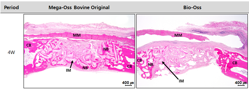
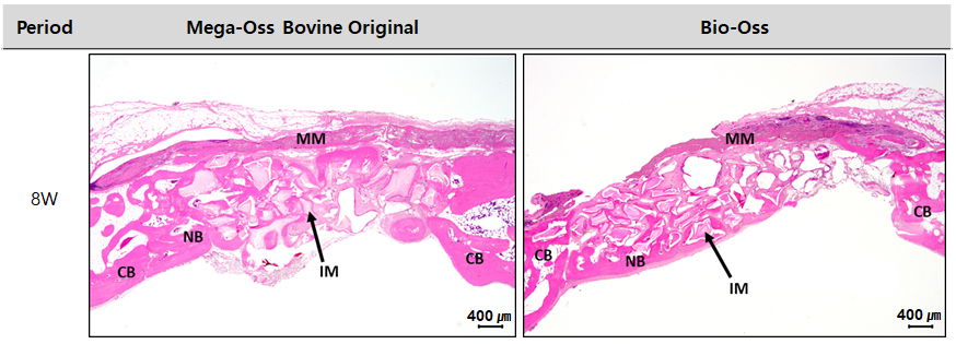
At 4 weeks after implantation, both the Mega-Oss Bovine Original and Bio-Oss groups showed minimal inflammatory responses (Table 5, Figs. 14 and 15). Slight fibrosis and scattered infiltration of a small number of granulocytes and lymphocytes were observed within the connective tissue between graft particles, particularly near the defect surface, suggesting normal postoperative tissue healing rather than a direct reaction to the graft materials. A few macrophages and multinucleated giant cells were observed adjacent to the graft particles in both groups, indicating a mild foreign body response. Mild connective tissue proliferation and limited neovascularization were also present between graft particles. No remarkable histological differences were identified between the two groups.
Table 5.
Evaluation of inflammation and tissue responses in the implantation areas on week 4 and 8 after the surgery
| Period | 4W | 8W | ||||||||||
| Group | Mega-Oss Bovine Original | Bio-Oss | Mega-Oss Bovine Original | Bio-Oss | ||||||||
| Animal ID | #1101 | #1102 | #1103 | #1101 | #1102 | #1103 | #1201 | #1202 | #1203 | #1201 | #1202 | #1203 |
|
Inflammatory cell infiltration around the implants* | 1+ | 1+ | 1+ | +/- | 1+ | 1+ | - | - | - | 1 | 2 | 1 |
| Cell type/response** | ||||||||||||
| PMN cells | 1 | 0 | 0 | 0 | 0 | 1 | 0 | 0 | 0 | 0 | 0 | 0 |
| Lymphocytes | 2 | 1 | 1 | 1 | 1 | 1 | 0 | 0 | 0 | 1 | 1 | 0 |
| Plasma cells | 0 | 0 | 0 | 0 | 0 | 0 | 1 | 0 | 1 | 0 | 1 | 0 |
| Macrophages | 1 | 1 | 1 | 1 | 1 | 1 | 0 | 0 | 0 | 1 | 2 | 1 |
| Giant cells | 0 | 1 | 1 | 1 | 1 | 1 | 1 | 1 | 1 | 2 | 1 | 2 |
| Necrosis | 0 | 0 | 0 | 0 | 0 | 0 | 0 | 0 | 0 | 0 | 0 | 0 |
| Sub-total (X2) | 8 | 6 | 6 | 6 | 6 | 8 | 4 | 2 | 4 | 8 | 10 | 6 |
| 6.7 ± 1.55 | 6.7 ± 1.55 | 3.3 ± 1.15 | 8.0 ± 2.00 | |||||||||
| Response** | ||||||||||||
| Neovascularization | 1 | 1 | 1 | 1 | 1 | 1 | 2 | 2 | 2 | 2 | 2 | 2 |
| Fibrosis | 2 | 2 | 2 | 2 | 2 | 2 | 2 | 2 | 2 | 2 | 2 | 2 |
| Fatty infiltration | 0 | 0 | 0 | 0 | 0 | 0 | 0 | 0 | 0 | 0 | 0 | 0 |
| Sub-total | 3 | 3 | 3 | 3 | 3 | 3 | 4 | 4 | 4 | 4 | 4 | 4 |
| 3.0 ± 0.00 | 3.0 ± 0.00 | 4.0 ± 0.00 | 4.0 ± 0.00 | |||||||||
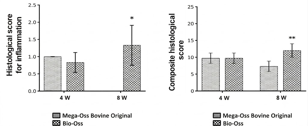
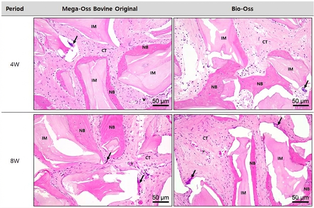
At 8 weeks after implantation, inflammatory cell infiltration was rarely observed in either group. Occasional macrophages and multinucleated giant cells remained adjacent to residual graft particles, although these cells appeared more frequently in the Bio-Oss group than in the Mega-Oss Bovine Original group. In both groups, connective tissue maturation progressed with increased fibroblast density and neovascularization compared with the 4-week observations.
Overall, both graft materials demonstrated favorable tissue compatibility with minimal inflammatory responses throughout the observation period. Although both groups showed mild cellular reactions to residual graft particles, the Mega-Oss Bovine Original group exhibited slightly less pronounced macrophage and multinucleated giant cell responses than the Bio-Oss group at 8 weeks post-implantation. Neovascularization and fibrosis were considered part of the normal tissue repair process rather than adverse reactions to the implanted graft materials.
Quantitative histomorphometric analysis was performed to evaluate newly formed bone and residual graft material within the defect area at 4 and 8 weeks post-implantation (Table 6, Figs. 16 and 17). At 4 weeks, the mean area of newly formed bone in the Mega-Oss Bovine Original group was 2.30 ± 0.91 mm2, corresponding to 23.4 ± 6.07% of the total defect area (9.64 ± 1.45 mm2). In the Bio-Oss group, the mean area of newly formed bone was 1.52 ± 0.44 mm2, corresponding to 18.0 ± 4.23% of the total defect area (8.46 ± 0.77 mm2). The mean area of residual graft material was 2.18 ± 0.50 mm2 in the Mega-Oss Bovine Original group and 1.35 ± 0.51 mm2 in the Bio-Oss group.
Table 6.
The areas of remnant implant materials and new bone in the cranial defective area
| Period | Mega-Oss Bovine Original | Bio-Oss | ||||||
| ID |
Remnant Implants (mm2) |
New bone (mm2) | % of new bone | ID |
Remnant Implants (mm2) |
New bone (mm2) | % of new bone | |
| 4W | #1101 | 2.18 | 1.5 | 17 | #1101 | 1.47 | 2.01 | 21.9 |
| #1102 | 2.68 | 3.29 | 29.1 | #1102 | 0.79 | 1.42 | 18.5 | |
| #1103 | 1.68 | 2.11 | 24 | #1103 | 1.78 | 1.14 | 13.5 | |
| Mean ± SD | 2.18 ± 0.50 | 2.3 ± 0.91 | 23.4 ± 6.07 | Mean ± SD | 1.35 ± 0.51 | 1.52 ± 0.44 | 18.0 ± 4.23 | |
| 8W | #1201 | 1.56 | 2.34 | 32.1 | #1201 | 0.37 | 1.72 | 16.3 |
| #1202 | 0.79 | 1.25 | 17.6 | #1202 | 0.61 | 1.7 | 22.2 | |
| #1203 | 3.5 | 4.94 | 28.1 | #1203 | 1.33 | 2.31 | 23 | |
| Mean ± SD | 1.95 ± 1.40 | 2.84 ± 1.90 | 25.93 ± 7.49 | Mean ± SD | 0.77 ± 0.50 | 1.91 ± 0.35 | 20.5 ± 3.66 | |
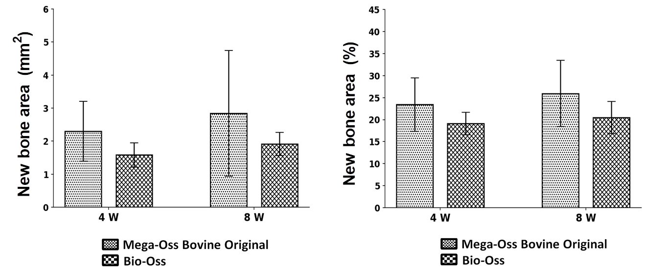
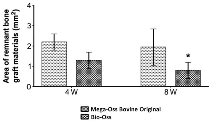
At 8 weeks, newly formed bone was observed throughout the defect area in both groups. The mean new bone area was 2.84 ± 1.90 mm2 in the Mega-Oss Bovine Original group and 1.91 ± 0.35 mm2 in the Bio-Oss group, corresponding to 25.93 ± 7.49% and 20.5 ± 3.66% of the total defect area, respectively. The mean area of residual graft material was 1.95 ± 1.40 mm2 in the Mega-Oss Bovine Original group and 0.77 ± 0.50 mm2 in the Bio-Oss group.
Although numerical differences were observed between the groups for both newly formed bone and residual graft area at each observation period, no statistically significant differences were identified between the Bio-Oss and Mega-Oss Bovine Original groups (p > .05). These findings indicate comparable patterns of bone formation and graft retention between the two materials during the observation period.
Ⅳ. Discussion
DBBM is one of the most extensively studied and clinically validated xenogeneic bone graft materials.20,21,22 In particular, Bio-Oss, produced through chemical treatment and low-temperature processing to remove organic components from bovine bone, is widely recognized as a benchmark material with well-documented biocompatibility and osteoconductive properties.23 These processing methods are known to influence the degradation behavior, bone formation, and overall biological performance of graft materials.24
In this study, the physicochemical properties, biocompatibility, and in vivo behavior of a novel xenograft, Mega-Oss Bovine Original, were systematically compared with those of Bio-Oss.
The pore structure of bone graft materials is a critical determinant of osteoconduction. Macropores (>100 μm) facilitate cellular infiltration and vascularization, whereas micropores (<10 μm) and mesopores (10–100 μm) enhance capillary-driven transport of blood and growth factors into the graft matrix. SEM observations in this study confirmed that both materials possessed well-developed macro-, micro-, and mesoporous structures. In particular, the distribution and morphology of micro- and mesopores in Mega-Oss Bovine Original were comparable to those of Bio-Oss, suggesting a similar microstructural environment conducive to bone regeneration.
Quantitative analysis further revealed that Mega-Oss Bovine Original exhibited a higher porosity (86.65%) and a larger specific surface area (127.015 m2/g) than Bio-Oss. These features are likely to enhance protein adsorption and blood wettability, thereby improving handling properties and promoting early osteoblast attachment. Such a biomimetic microenvironment is advantageous for initiating bone regeneration.
Crystallographic analysis using XRD and TEM demonstrated that both materials consisted of low-crystalline HA with nanocrystal sizes in the range of approximately 10–30 nm. This nanoscale, low-crystalline structure closely resembles the mineral phase of natural bone. Furthermore, lower crystallinity is generally associated with increased biodegradability and resorption rates, suggesting that these materials may facilitate gradual replacement by newly formed bone during the remodeling process.
FT-IR analysis confirmed that both materials shared similar chemical compositions, characterized by phosphate (PO43‒) and carbonate (CO32‒) groups typical of carbonated HA. The absence of distinct amide bands further indicated effective removal of organic components. The weak or indistinct hydroxyl (O–H) bands, commonly observed in highly crystalline HA, were consistent with the low-crystalline nature of both materials, corroborating the XRD and TEM findings.
For xenogeneic graft materials, thorough removal of organic components is essential to minimize potential immunological reactions. TGA results showed minimal weight loss in the temperature range associated with organic decomposition, indicating negligible residual organic content. This finding was further supported by Soxhlet and Kjeldahl analyses, which demonstrated very low levels of residual lipids and proteins in both Mega-Oss Bovine Original and Bio-Oss. The measured values were comparable between the two materials, suggesting that the purification processes effectively removed organic constituents that could potentially contribute to inflammatory or immunological reactions after implantation.
The in vitro cytotoxicity assessment using L-929 fibroblasts further confirmed the excellent biocompatibility of both materials, with cell viability values well above the ISO 10993-5 threshold. This indicates that the low residual organic content and high physicochemical purity do not adversely affect cellular responses, supporting their suitability for clinical application.
In vivo rabbit calvarial defect model, the present study evaluated bone regeneration and graft behavior using micro-CT, histological analysis, and histomorphometric measurements at 4 and 8 weeks post- implantation. Because different animals were used at each time point, the findings represent cross-sectional comparisons rather than longitudinal changes, and therefore should be interpreted as intergroup differences at specific healing stages.
Micro-CT analysis demonstrated mineralized tissue formation and the presence of residual graft materials in all defect sites at both time points. Statistical analysis revealed no significant differences between the Bio-Oss and Mega-Oss Bovine Original groups in terms of bone volume fraction (BV/TV) and bone mineral density (BMD) at either 4 or 8 weeks (p > .05). In particular, the BV/TV values at 8 weeks were nearly identical between the groups, indicating similar levels of mineralized tissue formation. Likewise, no significant difference was observed in BMD, suggesting that both materials maintained comparable mineralized tissue density throughout the healing period. Although the Mega-Oss Bovine Original group exhibited relatively larger standard deviations for BV/TV and BMD at 8 weeks, indicating a certain degree of inter-animal variability in the healing response, the overall pattern of bone regeneration was consistent with that observed in the Bio-Oss group. Collectively, these findings suggest that Mega-Oss Bovine Original provides osteoconductive performance comparable to that of Bio-Oss under the conditions of the present study. However, it should be noted that a technical limitation exists in the micro-CT analysis regarding the clear separation of newly formed bone from residual graft materials. Since both Bio-Oss and Mega-Oss Bovine Original exhibit mineral densities highly comparable to those of mineralized bone, the BV/TV values reported in this study represent the sum of both new bone and the remaining graft particles. Therefore, these findings should be interpreted in conjunction with the histomorphometric results for a more comprehensive evaluation of bone regeneration.
Histological evaluation demonstrated favorable tissue compatibility for both graft materials throughout the observation period. Inflammatory cell infiltration was minimal at 4 weeks, and at 8 weeks, it was rarely observed in the Mega-Oss Bovine Original group, whereas mild inflammatory cell infiltration was observed in the Bio-Oss group. Occasional macrophages and multinucleated giant cells were observed adjacent to residual graft particles, which is a commonly reported host response to implanted biomaterials and is generally associated with physiological tissue remodeling rather than adverse inflammatory reactions. In addition, connective tissue maturation and neovascularization were observed in both groups, suggesting ongoing tissue integration and repair within the defect area. Overall, the histological findings indicate that both Mega-Oss Bovine Original and Bio-Oss exhibited favorable tissue compatibility and comparable biological responses under the conditions of the present study.
Histomorphometric analysis further supported the findings obtained from the micro-CT and histological evaluations. Quantitative measurements of newly formed bone area and residual graft area showed no statistically significant differences between the Bio-Oss and Mega-Oss Bovine Original groups at either 4 or 8 weeks (p > .05). Although the Mega-Oss Bovine Original group exhibited numerically higher mean values for newly formed bone formation, particularly at 8 weeks, substantial inter-animal variability was also observed. Therefore, these numerical differences should be interpreted with caution and are not sufficient to demonstrate a meaningful difference in regenerative performance between the two materials. Rather, the overall histomorphometric findings suggest that both materials supported comparable levels of new bone formation during the healing period.
With regard to residual graft behavior, deproteinized bovine bone mineral (DBBM) materials such as Bio-Oss are known to exhibit slow resorption and prolonged persistence after implantation. Previous histological studies have demonstrated that Bio-Oss particles may remain detectable in human biopsy specimens for several years following grafting procedures. Therefore, the differences in residual graft area observed in the present study should not be interpreted as evidence of different resorption characteristics between the two materials. Instead, these findings are more likely attributable to biological variability among animals, sampling differences, and the relatively short observation period. Consequently, longer-term studies are required to further evaluate graft remodeling and residual graft behavior over time.25,26
Ⅴ. Conclusion
In this study, the physicochemical properties and in vivo bone regeneration capacity of a newly developed xenogeneic bone graft material, Mega-Oss Bovine Original, were evaluated in comparison with Bio-Oss. Mega-Oss Bovine Original exhibited a low-crystalline hydroxyapatite structure, and differences were observed in porosity and specific surface area compared with the control material. Other physicochemical parameters, including thermal stability and residual organic content, were comparable between the two materials, indicating overall material consistency. In the rabbit calvarial defect model, greater new bone formation was observed in the Mega-Oss Bovine Original group at 8 weeks compared with Bio-Oss. This finding represents an intergroup difference at a specific healing stage, and may be associated with the physicochemical characteristics of the material, although causal relationships cannot be confirmed based on the present study design. Taken together, these results suggest that Mega-Oss Bovine Original is a biocompatible xenogeneic bone graft material that supports new bone formation in a rabbit calvarial defect model. Further long-term and adequately powered studies are required to clarify its comparative performance and clinical relevance