


Ⅰ. Introduction
Ⅱ. Case Report
1. Case 1
2. Case 2
3. Clinical assessment
4. Radiographic assessment
Ⅲ. Discussion
Ⅰ. Introduction
Periodontitis is a leading cause of tooth extraction, ranging from 24.8% to 38.1% of extracted teeth in adults.1 It often presents as a generalized condition across the entire dentition rather than being localized to a single site.2 Consequently, even when periodontally compromised teeth are extracted, residual periodontal issues may persist in adjacent areas. These compromised sites pose significant challenges during implant placement. Because the alveolar crest of the adjacent teeth remains unchanged after extraction, it may require a deeper implant placement, potentially leading to increased peri-implant bone loss.3 Additionally, such sites may serve as reservoirs for microbial transmission, increasing the risk of peri-implantitis and implant failure.4 Therefore, ensuring a stable and healthy periodontal and peri-implant environment adjacent to the planned implant site before implant placement is crucial for the long-term success of implant therapy.
Non-surgical periodontal therapy, including scaling and root planing, has been shown to reduce bacterial load.5 However, residual intrabony or peri-implant defects may persist despite initial therapy, requiring surgical intervention such as open flap debridement, or regenerative or resective procedures.6 Regenerative therapy is widely used for periodontal intrabony defects and has been shown to promote long-term tooth retention and improve clinical parameters.7 Similarly, regenerative approaches have been applied to peri-implant defects to restore lost supporting bone and improve peri-implant tissue stability.8
Conventionally, regenerative treatment of these compromised sites is completed before implant placement. Although this staged approach allows adequate healing before implant placement, it requires additional surgical procedures, which may contribute to further bone loss, increased patient discomfort, and prolonged treatment time.9 Consequently, simultaneous implant placement with bone grafting has been proposed as a viable approach for restoring compromised sites.10 However, to the best of our knowledge, little information is available regarding the simultaneous surgical management of compromised teeth or implants adjacent to planned implant sites at the time of implant placement.
This case report presents two cases in which implant placement was performed simultaneously with the surgical treatment of compromised adjacent sites. One case involved a residual periodontal intrabony defect adjacent to the planned implant site, whereas the other involved a peri-implant defect adjacent to the implant placement site. The objective of this case report is to describe the 3-year clinical and radiographic outcomes of this simultaneous treatment approach.
Ⅱ. Case Report
This case report was conducted with the approval of the Institutional Review Board of Seoul National University Dental Hospital (ERI25017) and was prepared in accordance with the CARE guidelines.11
1. Case 1
A 70-year-old non-smoking female with hypertension (ASA-II according to the American Society of Anesthesiologists Physical Status Classification System)12 was referred from the Department of Conservative Dentistry for the evaluation of the left mandibular first molar (tooth #36 in the FDI system). She presented with swelling and bleeding during tooth brushing in the left mandibular posterior region. Clinical examination revealed a buccal sinus tract and pus discharge from tooth #36. A periapical radiograph showed severe bone loss, and extraction was planned. Inflammatory signs were also observed in the left mandibular posterior region, prompting non-surgical periodontal treatment.
Four months after tooth extraction, implant placement was planned. A panoramic radiograph taken before implant placement showed unresolved bone loss on the distal aspect of the left mandibular second premolar (tooth #35 in the FDI system) and the absence of tooth #36 (Fig. 1). Preoperative clinical photographs revealed slight gingival recession on the distal aspect of tooth #35 (Fig. 2A and 2B).
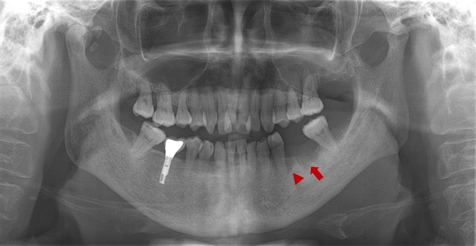
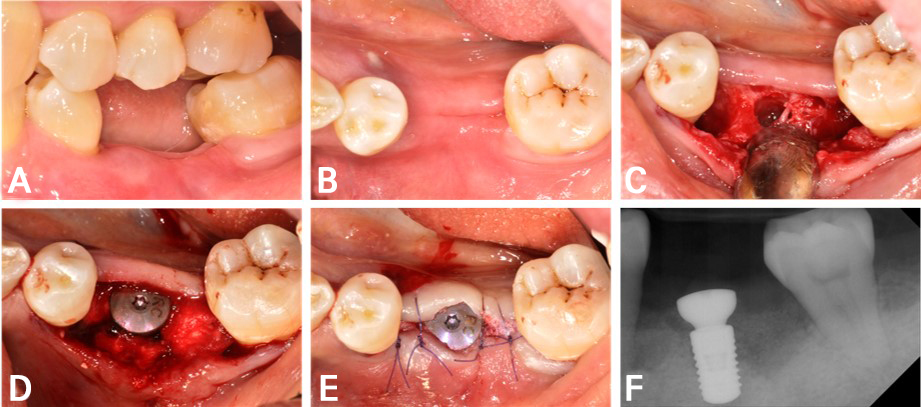
Fig. 2.
Surgical procedure in Case 1. (A) Preoperative clinical photograph showing the edentulous site #36 and the distal aspect of tooth #35, (B) Occlusal view of the edentulous ridge before surgery, (C) Following flap elevation, an intrabony defect was identified on the distal aspect of tooth #35 after debridement of inflammatory tissue and root planing, (D) An implant was placed at site #36, and bone grafting with collagenated deproteinized bovine bone mineral was performed at both the implant site and the adjacent intrabony defect, (E) Flap closure was achieved with simple interrupted monofilament sutures, (F) Immediate postoperative periapical radiograph confirming appropriate implant positioning and graft placement.
The implant was placed simultaneously with the treatment of the residual periodontal defect adjacent to the planned implant site. Under local anesthesia, a lingually positioned crestal incision was made without vertical releasing incisions. A full-thickness flap was then elevated, exposing a 3-wall intrabony defect with a vertical depth of 10 mm on the distal aspect of tooth #35. After removal of the granulation tissue and root planing, no chemical treatment or root surface conditioning was performed on the adjacent tooth. An osteotomy was performed at site #36 for implant placement (Fig. 2C). An implant (BL Straumann Ø4.8 × 8 mm; Institute Straumann AG, Basel, Switzerland) was placed in the #36 site, and bone grafting was performed using collagenated deproteinized bovine bone mineral (BioOss-Collagen; Geistlich Pharma AG, Wolhusen, Switzerland) at both the implant site and the intrabony defect of tooth #35 (Fig. 2D). A healing abutment (RC Healing abutment Ø6.5 × 4.0 mm; Institute Straumann AG) was connected, and a collagen sponge (Teruplug; Terumo Co., Tokyo, Japan) was applied over the graft material. Simple interrupted sutures were placed using a monofilament suture (Monosyn 5/0; B Braun Aesculap, Tuttlingen, Germany) (Fig. 2E).
Postoperatively, the patient was prescribed antibiotics (100 mg cefdinir; Jeil Pharmaceutical, Seoul, Korea) and an analgesic (650 mg acetaminophen; Hanmi Pharmaceutical, Seoul, Korea) three times daily for 7 days. The patient was also instructed to rinse with a 0.2% chlorhexidine solution twice daily for 1 minute for 7 days.
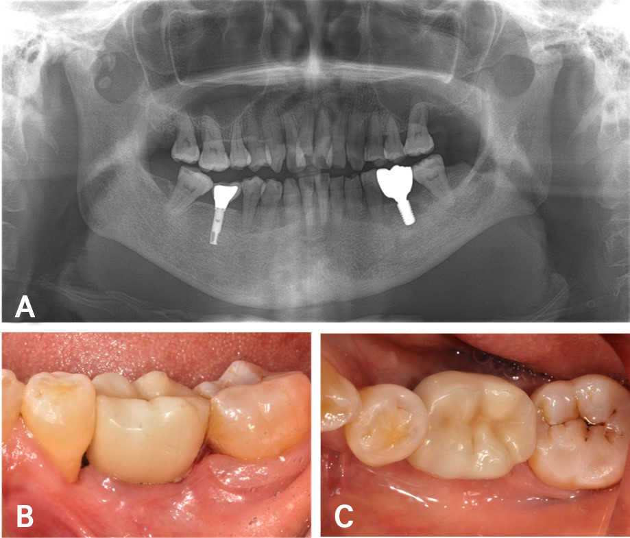
One week after surgery, the sutures were removed.Radiographic evaluation showed the implant fixture in place with visible bone graft material (Fig. 2F). An impression for fabrication of the definitive prosthesis was taken 4 months after surgery, and the prosthesis was connected to the implant 5 months postoperatively (Fig. 3).
Three years after surgery, panoramic and periapical radiographs showed bone fill on the distal aspect of tooth #35, and the implant at site #36 remained stable without peri-implant bone loss (Fig. 4A and 4B). Clinical examination also confirmed the absence of inflammatory signs, demonstrating stable peri-implant tissue conditions (Fig. 4C and 4D).
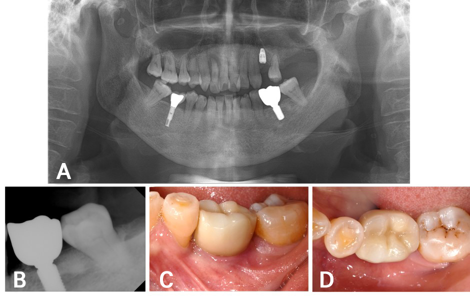
Fig. 4.
Three-year follow-up after implant placement. (A, B) Panoramic and periapical radiographs obtained 3 years after surgery demonstrating stable peri-implant bone levels, (C, D) Clinical photographs obtained at the 3-year follow-up showing healthy peri-implant and adjacent periodontal tissues with no clinical signs of inflammation.
2. Case 2
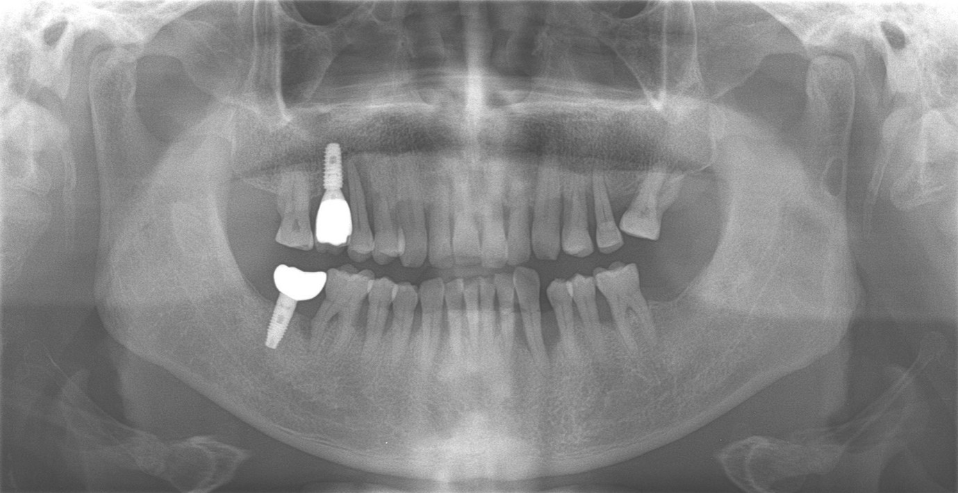
A 69-year-old male patient, who smoked approximately two-thirds of a pack of cigarettes per day with no systemic diseases (ASA-II; according to the American Society of Anesthesiologists Physical Status Classification System),12 presented with severe mobility and discomfort associated with the maxillary right second premolar (tooth #15 in the FDI system). A panoramic radiograph revealed extensive bone loss around tooth #15 extending toward the implant at the maxillary right first molar site (implant #16 in the FDI system) (Fig. 5). Clinical examination confirmed vertical mobility of tooth #15. The tooth was considered as hopeless, and was extracted.
Four months after tooth extraction, implant placement was performed. A periapical radiograph taken after extraction revealed vertical bone loss at site #15 and on the mesial aspect of implant #16 (Fig. 6A). Preoperative intraoral photographs showed slight soft tissue recession at site #15 and gingival recession on the distal aspect of the maxillary right first premolar (tooth #14 in the FDI system) (Fig. 6B and 6C).
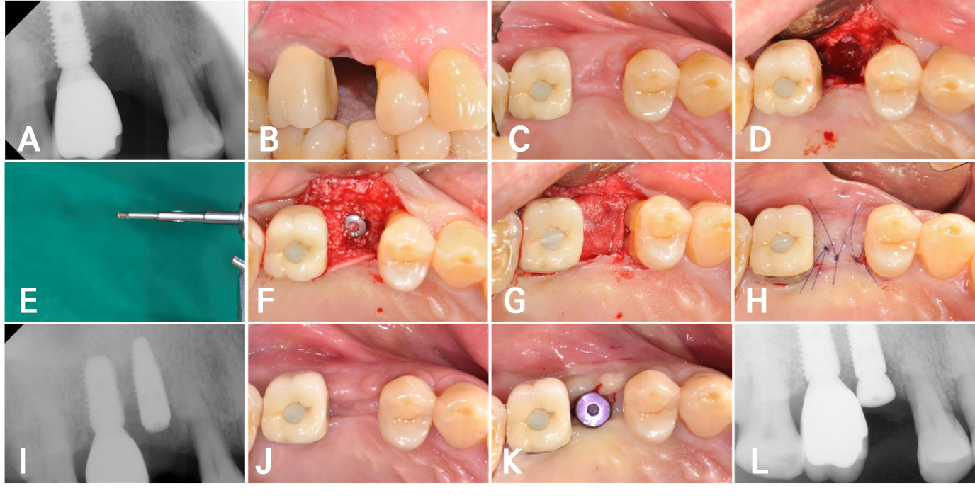
Fig. 6.
Surgical procedure in Case 2. (A) Preoperative periapical radiograph showing bone loss at site #15 extending to the mesial aspect of implant #16, (B, C) Preoperative clinical photographs showing the edentulous ridge and soft tissue condition around site #15 and implant #16, (D) Following flap elevation, an intrabony defect extending from site #15 to the mesial aspect of implant #16 was identified, (E) Titanium brush was used for implant surface decontamination, (F) An implant was placed at site #15 and a cover screw was connected, followed by bone grafting with collagenated deproteinized bovine bone mineral at the implant site and the mesial peri-implant defect, (G) A double layer of resorbable collagen membrane was placed over the grafted area, (H) Flap closure was achieved with simple interrupted monofilament sutures, (I) Immediate postoperative periapical radiograph confirming appropriate implant positioning and graft placement, (J) Clinical photograph obtained 3 months after surgery showing favorable soft tissue healing, (K) A healing abutment was connected following a short crestal incision, (L) Periapical radiograph confirming appropriate connection of the healing abutment and maintenance of the grafted area.
A simultaneous surgical approach was used to treat the peri-implant defect adjacent to the planned implant site and place the implant during the same surgical procedure. Following local anesthesia, a crestal incision positioned palatal to the ridge crest was made without vertical releasing incisions. A full-thickness flap was then elevated, revealing a continuous 3-wall intrabony defect (Class 1b) with a vertical depth of 9 mm extending from site #15 to the mesial aspect of implant #16 (Fig. 6D).13 Granulation tissue at edentulous site #15 was debrided, and implant #16 was mechanically decontaminated using a titanium brush (TN-Brush; Dentium, Suwon, Korea) (Fig. 6E), followed by application of hydrogen peroxide (H₂O₂) with a soaked cotton pellet and doxycycline irrigation. An implant (BLT Straumann Ø4.1 × 12 mm; Institute Straumann AG) was placed at site #15, and a cover screw (RC closure cap Ø3.5 × 0.5 mm; Institute Straumann AG) was connected. Bone grafting was carried out using collagenated deproteinized bovine bone mineral (BioOss-Collagen; Geistlich Pharma AG) to fill the bony defect (Fig. 6F). A resorbable membrane (Bio-Gide; Geistlich Pharma) was placed as a double layer over the grafted site (Fig. 6G), followed by simple interrupted suturing with a monofilament suture (Monosyn 5/0; B Braun Aesculap) (Fig. 6H).
After the operation, a regimen of antibiotics (100 mg cefdinir; Jeil Pharmaceutical, Seoul, Korea) and analgesic (650 mg acetaminophen; Hanmi Pharmaceutical) was administered three times a day for 5 days. The patient was also instructed to rinse with a 0.2% chlorhexidine solution twice daily for 1 week.
One week after surgery, the sutures were removed, and a radiograph confirmed the presence of the implant along with the grafted bone material (Fig. 6I). Three months after implant placement, the site exhibited proper healing (Fig. 6J). A short crestal incision was made, and the cover screw was replaced with a healing abutment (RC Healing abutment Ø5.0 × 4.0 mm; Institute Straumann AG) (Fig. 6K). The same antibiotic and analgesic regimen prescribed after implant placement was administered for 5 days. A periapical radiograph demonstrated the healing abutment in place and increased radiopacity of the grafted area (Fig. 6L). 4 months after surgery, a definitive impression was obtained for prosthesis fabrication. The definitive prosthesis was attached to the implant 1 month later, and a radiographic evaluation was performed (Fig. 7).
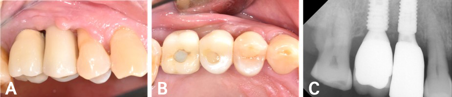
Fig. 7.
Prosthetic rehabilitation 5 months after surgery. (A, B) Clinical photographs obtained 5 months after surgery, showing the definitive implant-supported prosthesis connected to the implant, (C) Periapical radiograph demonstrating stable peri-implant bone conditions after prosthetic rehabilitation.
Three years after surgery, panoramic and periapical radiographs confirmed stable bone levels around implants #15 and #16 (Fig. 8A and 8B). Although a slight increase in gingival recession was noted on the distal aspect of tooth #14, no signs of inflammation were observed (Fig. 8C and 8D).
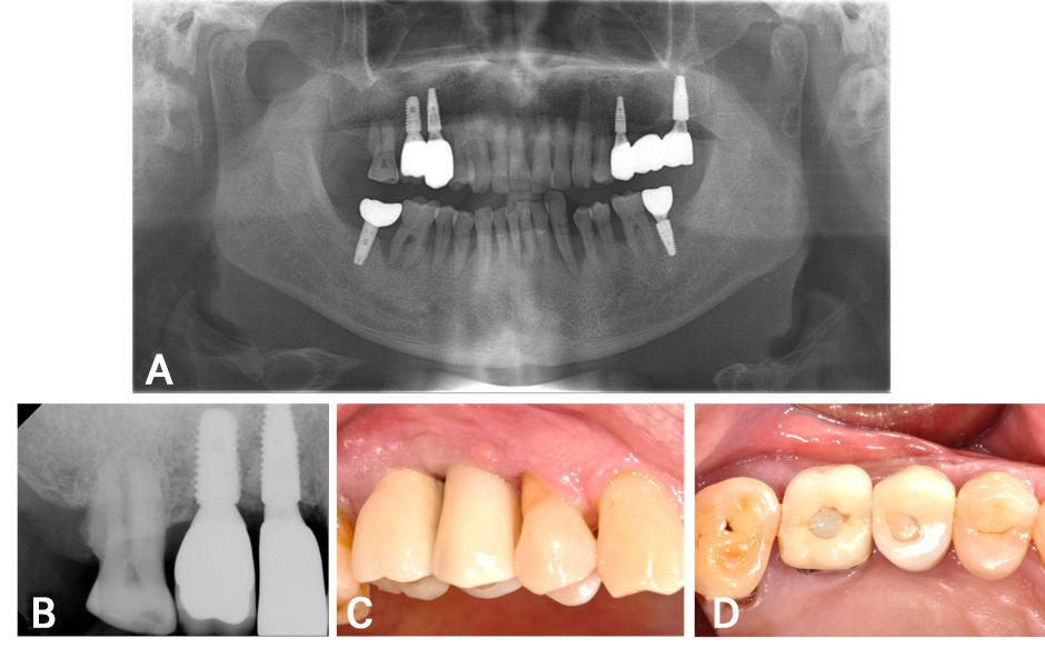
Fig. 8.
Three-year follow-up after implant placement. (A, B) Panoramic and periapical radiographs obtained 3 years after surgery demonstrating stable peri-implant bone levels, (C, D) Clinical photographs obtained at the 3-year follow-up showing healthy peri-implant soft tissues and a stable prosthetic restoration with no clinical signs of inflammation.
3. Clinical assessment
The following clinical parameters were measured at the study sites at baseline (pre-surgery) and at the 3-year follow-up.
• Probing pocket depth (PPD) was measured at six sites per tooth/implant (mesiobuccal, midbuccal, distobuccal, mesiolingual, midlingual, and distolingual) using a periodontal probe (Hu-Friedy CP-12/Thin Williams Color Coded Probe; Hu-Friedy, Chicago, USA).14 Recession (REC) was assessed as the distance from the cemento-enamel junction (CEJ) or restorative margin to the free gingival or mucosal margin at the same six sites used for PPD measurement. For the changes between baseline and the 3-year follow-up, a negative REC value indicated increased gingival recession.15 Keratinized gingival/mucosal width (KGW/KMW) was evaluated as the distance from the free gingival or mucosal margin to the mucogingival junction, measured at the midbuccal site.16 When calculating the difference between baseline and the 3-year follow-up, a negative value indicated a decrease in the width of keratinized gingiva/mucosa.
• Vestibular depth (VD) was determined as the distance from the mucosal margin to the most concave point of the mucosal fold at the midbuccal site.17
• The plaque index (PI) and sulcus bleeding index (SBI) were recorded around teeth,18,19 whereas the modified plaque index (mPI) and modified sulcus bleeding index (mSBI) were assessed around implants.20
• The full mouth plaque index (FMPI) was calculated as the proportion of tooth surfaces with plaque relative to the total number of tooth surfaces examined.21
4. Radiographic assessment
Radiographic bone level (RBL) was evaluated at the mesial and distal sites using periapical and panoramic radiographs.22 For teeth, RBL was measured as the distance from the CEJ to the crestal bone; for implants, it was measured from the implant platform to the crestal bone. A negative RBL indicated that the bone was positioned coronal to the implant shoulder. Changes observed between baseline and the 3-year follow-up indicate the extent of bone gain.
The clinical parameters for each patient at baseline and at the 3-year follow-up are presented in Table 1.
Table 1.
Clinical parameters at the baseline and 3-year follow-up
3y FU, 3-year follow-up; PPD, probing pocket depth; REC, recession; KGW/KMW, keratinized gingival/mucosal width; VD, vestibular depth; PI, plaque index; mPI, modified plaque index; SBI, sulcus bleeding index; mSBI, modified sulcus bleeding index; FMPI, full mouth plaque index; RBL, radiographic bone level; PPD and REC, measured at six sites of the tooth/implant. MB, mesiobuccal; B, midbuccal; DB, distobuccal; ML, mesolingual; L, midlingual; DL, distolingual; M, mesial; D, distal.
In Case 1, for tooth #35, which underwent bone grafting, the mean PPD had decreased by 2.5 mm, while the mean REC had increased by 0.8 mm at the 3-year follow-up. KGW/KMW decreased by 2.0 mm, whereas VD remained unchanged. PI remained unchanged, SBI decreased by 1, and FMPI decreased from 25.8% to 16.7% (a decrease of 9.1 percentage points). RBL assessment indicated a mean bone fill of 3.9 mm.
For implant #36, which was placed simultaneously with bone grafting, PPD remained ≤3.0 mm, and no gingival recession was observed at the 3-year follow-up. KGW/KMW was 4.0 mm, and VD was 6.0 mm. The mPI was 1, and the mSBI was 0. Radiographic evaluation revealed an RBL of -0.7 mm relative to the implant shoulder.
In Case 2, for implant #16, which underwent regenerative therapy, the mean PPD had decreased by 1.8 mm, with no gingival recession. KGW/KMW increased by 1.0 mm, whereas VD decreased by 2.0 mm. No changes were noted in mPI or mSBI, while FMPI decreased by 11.62%. RBL assessment revealed a mean bone fill of 4.1 mm.
For implant #15, which was placed simultaneously with bone grafting, PPD remained ≤4.0 mm and no gingival recession was observed at the 3-year follow-up. KGW/KMW was 3.0 mm, and VD was 6.0 mm. Both mPI and mSBI were 1, and radiographic analysis indicated an RBL of -0.7 mm relative to the implant shoulder.
Ⅲ. Discussion
The present report highlights the feasibility of simultaneously treating compromised periodontal or peri-implant lesions adjacent to planned implant sites during implant placement. Despite differences in the adjacent defects, both patients demonstrated favorable clinical and radiographic outcomes that were maintained throughout the 3-year follow-up period. These findings suggest that simultaneous management of adjacent compromised sites may reduce the need for staged surgical procedures while maintaining stable periodontal and peri-implant conditions in carefully selected patients.
The management of compromised tissues adjacent to planned implant sites presents both biological and clinical challenges. Residual periodontal or peri-implant inflammation may serve as a source of microbial contamination and compromise peri-implant tissue stability after implant placement. Therefore, successful treatment requires not only replacement of the missing tooth but also appropriate management of adjacent periodontal or peri-implant lesions. Previous preclinical evidence has suggested that vertical bone augmentation can promote periodontal regeneration in neighboring teeth.23
Conventionally, compromised adjacent teeth or implants are treated before implant placement using a staged approach. Although this strategy provides adequate infection control and healing time, it inevitably increases treatment duration and requires an additional surgical intervention. In contrast, the present cases demonstrate that simultaneous surgical management of adjacent lesions and implant placement can be successfully performed within a single procedure, potentially reducing patient morbidity while maintaining favorable clinical outcomes. In Case 2, a titanium brush was used for implant surface decontamination, which is supported by previous studies demonstrating effective biofilm removal while preserving implant surface characteristics.24,25,26
Regenerative treatment outcomes may vary according to defect morphology. In the present report, both cases involved deep 3-wall intrabony defects. However, in Case 1, the intrabony defect was confined to the adjacent tooth, whereas in Case 2, the peri-implant intrabony defect extended into the site of the newly placed implant, resulting in a wider defect. Previous studies have shown that deep, narrow intrabony defects generally exhibit greater radiographic bone gain and clinical attachment level gain following periodontal regenerative therapy than wider defects.27,28 Accordingly, the wider defect in Case 2 might have been considered less favorable. Nevertheless, Cases 1 and 2 demonstrated comparable radiographic bone gains of 3.9 and 4.1 mm, respectively, as well as clinical attachment level gains of 1.7 and 1.8 mm, respectively. These comparable outcomes may be partly attributable to the 3-wall configuration of both defects. The remaining bony walls may have provided greater defect containment and stabilization of the graft material and blood clot, thereby creating a favorable environment for regenerative healing.
Flap design is an important determinant of wound stability and healing in regenerative procedures. In both cases, a crestal incision shifted toward the lingual or palatal aspect was made without vertical releasing incisions, followed by full-thickness flap elevation. Because adequate access to the defects was achieved through the crestal incision alone, vertical releasing incisions were omitted to minimize surgical trauma and preserve flap vascularity.29 In addition, the crestal incision was positioned lingually or palatally to keep the incision line away from the underlying defect and grafted area, thereby reducing the risk of wound dehiscence and graft exposure.30 Full-thickness flap elevation provided direct visualization and sufficient access for thorough removal of granulation tissue from the underlying defects.
Bone grafting was performed using collagenated deproteinized bovine bone mineral because of its favorable biocompatibility, slow resorption, and volume stability.31,32,33 A resorbable collagen membrane was additionally used in Case 2 according to the principles of guided bone regeneration to facilitate selective cell repopulation and space maintenance.34 Previous studies have consistently demonstrated favorable outcomes with these biomaterials in periodontal and peri-implant reconstructive procedures.35,36
Several healing protocols have been investigated following reconstructive treatment of peri-implantitis. Submerged healing has traditionally been advocated to protect the grafted site and promote undisturbed healing.37 However, this approach generally requires removal of the prosthesis before surgery, followed by a second-stage procedure and prosthetic reconnection. Therefore, transmucosal healing with the prosthesis maintained may be considered in carefully selected cases when adequate surgical access, implant surface decontamination, graft stability, and tension-free wound closure can be achieved. A retrospective study comparing prosthesis removal and retention following regenerative surgery found no significant difference in radiographic bone gain between the two approaches.38 Furthermore, a case series and a prospective cohort study demonstrated radiographic defect fill and favorable clinical outcomes after transmucosal reconstructive therapy performed without removal of the prosthetic restoration.39,40 Accordingly, in Case 2, the existing prosthesis was retained during reconstructive surgery to simplify the treatment procedure and avoid additional prosthetic and surgical interventions.
Oral hygiene is a well-established modifiable risk factor for both periodontal and peri-implant diseases. Poor plaque control has been associated with an increased risk of peri-implant mucositis, peri-implantitis, and compromised surgical outcomes.41,42,43 Both patients maintained favorable plaque control throughout the observation period, which likely contributed to the stable clinical and radiographic outcomes observed at the 3-year follow-up.
Long-term implant success depends not only on successful osseointegration but also on the maintenance of healthy peri-implant hard and soft tissues. Long-term studies have demonstrated favorable outcomes following regenerative treatment of periodontal intrabony defects,7,44 while similar reconstructive principles have also been successfully applied to peri-implant defects when adequate implant surface decontamination and favorable defect morphology are present.24,28,45 The present cases extend these concepts by demonstrating that treatment of adjacent compromised sites can be successfully integrated with implant placement during the same surgical procedure.
Several limitations should be acknowledged. First, this report includes only two cases, limiting generalization of the findings. Second, histologic confirmation of true periodontal regeneration or peri-implant bone regeneration was not available. Third, the surgical approach differed slightly according to the morphology of the adjacent defects; therefore, a standardized treatment protocol cannot be established from these cases. Finally, although the 3-year follow-up demonstrated stable outcomes, longer observation periods and controlled clinical studies are necessary to confirm the long-term predictability of this treatment strategy.
Within the limitations of this case report, simultaneous surgical treatment of compromised periodontal or peri-implant lesions adjacent to planned implant sites appears to be a feasible treatment strategy for selected patients. Careful case selection, meticulous surgical debridement, appropriate regenerative or reconstructive procedures, and supportive maintenance may contribute to favorable long-term peri-implant health.