


Ⅰ. Introduction
Assessing implant stability from a clinical perspective is critical for predicting the success of dental implant surgery. Osseointegration is defined as a direct functional and structural connection between the loaded implant surface and alveolar bone; in particular, primary stability immediately after implant placement is considered critical for achieving stable osseointegration and favorable clinical outcomes.1,2,3
As immediate implant placement and flapless surgery have become increasingly adopted to minimize patient discomfort and shorten treatment duration, the importance of objectively assessing implant stability has also increased.4,5In the posterior region, the irregular morphology of the residual alveolar bone following tooth extraction, combined with insufficient bone volume and heterogeneous bone density, may limit the bone–implant contact area, making primary stability particularly critical.
The flapless surgical technique offers advantages such as reduced postoperative pain and swelling, preservation of blood supply, and favorable soft tissue healing by avoiding flap elevation.6,7 However, limited surgical visibility remains a practical limitation.8 In this clinical setting, direct assessment of implant stability is challenging; therefore, devices that can objectively and noninvasively quantify implant stability are increasingly important.9 In clinical practice, implant stability assessment serves two distinct purposes: confirming primary stability at the time of placement and evaluating the progression of osseointegration before prosthetic restoration. The latter is particularly important because it provides an objective basis for determining the appropriate timing of prosthetic loading.
Osstell ISQ®, based on resonance frequency analysis (RFA), is widely regarded as the clinical standard for implant stability assessment because of its high reliability. However, each measurement requires removal of the healing abutment and attachment of a dedicated SmartPeg®, which is a practical limitation in clinical use.10,11 In flapless procedures, where preservation of peri-implant soft tissue healing is particularly important, repeated removal and reattachment of these components may cause minor mechanical trauma to the peri-implant soft tissues. Furthermore, when primary stability has not yet been sufficiently achieved, the torque generated during repeated component exchange may potentially cause microtrauma at the bone–implant interface.4
In contrast, AnyCheck®, which uses a damping capacity analysis (DCA) approach, enables percussion-based measurement without removal of the healing abutment, thereby reducing chair time. Compared with the conventional percussion device Periotest®, AnyCheck® reduces the number and intensity of percussions and incorporates an auto-stop function, thereby minimizing mechanical stress on peri-implant tissues.12,13
Although Osstell ISQ® is widely regarded as the clinical standard, its requirement for repeated healing abutment removal poses a particular limitation in flapless immediate implant placement, in which preservation of soft tissue is critical. This clinical gap raises the question of whether a percussion-based device that does not require abutment removal could serve as a reliable alternative. We therefore hypothesized that AnyCheck® would show clinically comparable agreement with Osstell ISQ® for assessing implant stability and that this agreement would increase as osseointegration progressed. The present study aimed to evaluate implant stability at implant placement and prosthetic impression in patients who underwent immediate implant placement following molar extraction using a flapless technique. Implant stability was assessed using Osstell ISQ® and AnyCheck®, yielding Implant Stability Quotient (ISQ) and Implant Stability Test (IST) values, respectively.
Ⅱ. Materials and Methods
This retrospective study included patients who underwent immediate single implant placement following molar extraction using a flapless technique at the Department of Oral and Maxillofacial Surgery, Inje University Sanggye Paik Hospital, between 2020 and 2022. The study was approved by the Institutional Review Board of Inje University Sanggye Paik Hospital (No. 2025-12-005). The requirement for individual informed consent was waived by the Institutional Review Board because of the retrospective nature of the study. A total of 45 patients (18 males and 27 females; mean age, 69.1 years) received 52 implants, including 24 maxillary and 28 mandibular implants. The indications for tooth extraction were periodontal disease (n = 18), dental caries (n = 14), residual roots (n = 9), and tooth fracture (n =11). The distribution of implant positions was as follows: #16 (n = 7), #17 (n = 5), #26 (n = 6), #27 (n = 6), #36 (n = 10), #37 (n = 5), #46 (n = 11), and #47 (n = 2).
Surgical procedures were performed using a flapless technique. Teeth were extracted atraumatically, and the extraction sockets were thoroughly debrided to remove granulation tissue before immediate implant placement. The implants used were sand-blasted and acid-etched surface bone-level implants (TS III SA; Osstem Implant Co., Busan, Korea; n = 31), sand-blasted and acid-etched surface tissue-level implants (SS III SA; Osstem Implant Co., n = 3), sand-blasted oxidized hydrophilic surface bone-level implants (TS III SOI; Osstem Implant Co., n = 2), sand-blasted and acid-etched surface bone-level implants (SimpleLine; Dentium Co., Seoul, Korea; n = 13), and clean sand-blasted and acid-etched surface bone-level implants (SQ; Dentis Co., Daegu, Korea; n = 3). Implant diameters were 4.5 mm (n = 2), 5.0 mm (n = 41), 5.5 mm (n = 8), and 6.0 mm (n = 1), and implant lengths were 7.0 mm (n = 7), 8.0 mm (n = 12), 8.5 mm (n = 23), and 10.0 mm (n = 10). Bone grafting was performed at 33 of the 52 implant sites using RenewOss (Renew Medical Co., Ltd., Bucheon, Korea; n = 20), DO Bone (Renew Medical Co., Ltd., n = 4), Inducera (Oscotec Inc., Seongnam, Korea; n = 3), InterOss (SigmaGraft Biomaterials, Brea, CA, USA; n = 3), CollaCera (Oscotec Inc., n = 1), or a combination of RenewOss and Inducera (n = 2). After implant placement, implants were allowed to heal before prosthetic impression taking. The mean interval between implant placement and impression taking was 2.8 months (84.3 ± 29.4 days). Implant placement was performed according to a standardized surgical protocol. Implant stability was assessed using two devices: Osstell ISQ® (Osstell AB, Gothenburg, Sweden) and AnyCheck® (Neobiotech, Wonju, Korea). Measurements were obtained at two time points: on the day of implant placement and on the day of prosthetic impression taking. The timing of prosthetic impression taking was determined based on the surgeon's clinical judgment, including assessment of the bone condition around the extraction socket and implant fixture and the intraoperative insertion torque, rather than on measured ISQ or IST values. Stability measurements were performed solely to assess osseointegration at the time of prosthetic impression taking.
For Osstell ISQ®, a SmartPeg® was connected to the implant, and measurements were obtained in four directions (mesial, buccal, distal, and lingual). The mean of the four measurements was used as the representative ISQ value. For AnyCheck®, a single measurement was obtained from the buccal aspect of the healing abutment. The device automatically delivered six consecutive taps and converted the resulting response into a single IST value. At each measurement session, ISQ was measured first, followed by IST. All measurements were performed by a single experienced surgeon.
Statistical analyses were performed to evaluate the correlation and clinical agreement between ISQ and IST values. Descriptive statistics are presented as means ± standard deviations (SDs) with 95% confidence intervals (CIs). Data normality was assessed using the Shapiro–Wilk test. Paired t-tests were used to compare ISQ and IST values between implant placement and prosthetic impression taking, and effect sizes were calculated using Cohen’s d with 95% CIs. Passing–Bablok regression analysis was performed to evaluate the relationship between ISQ and IST values at each time point. Agreement between the two devices was assessed using Bland–Altman analysis. Raw ISQ and IST values were used for this analysis, and the mean bias and 95% limits of agreement (LoA) were calculated. Categorical agreement was evaluated using a stability cutoff of ≥75.14,15,16 Percent agreement and Cohen’s κ were calculated at each time point, and κ values were interpreted according to the criteria of Landis and Koch (1977).17 All statistical analyses were performed using SPSS (version 25.0; IBM Corp., Armonk, NY, USA) and R (version 4.5.2; R Foundation for Statistical Computing, Vienna, Austria). Statistical significance was set at p < 0.05.
Ⅲ. Results
All 52 implants were clinically successful, with no implant failures or complications observed during the mean follow-up period of 42.0 ± 17.7 months after prosthetic restoration. ISQ and IST values were collected at both time points, with no missing data, and were included in the analysis.
ISQ values significantly increased from 76.0 ± 6.8 at implant placement to 81.8 ± 5.2 at prosthetic impression taking (paired t-test, p < .001; Cohen’s d = 1.09, 95% CI: 0.74–1.43). IST values also increased from 77.6 ± 6.0 to 82.5 ± 3.7 over the same period (p < .001; Cohen’s d = 0.75, 95% CI: 0.44–1.06). Both measurements showed large effect sizes. The SDs for both ISQ and IST values were lower at prosthetic impression taking than at implant placement (Table 1).
Table 1.
Changes in implant stability between implant placement and prosthetic impression measured by Osstell ISQ® and AnyCheck®
| Variable | Mean ± SD | Effect size† (95% CI) | p-value | |
| ISQ | Implant placement | 76.0 ± 6.8 | 1.09 (0.74–1.43) | < .001 |
| Prosthetic impression | 81.8 ± 5.2 | |||
| IST | Implant placement | 77.6 ± 6.0 | 0.75 (0.44–1.06) | < .001 |
| Prosthetic impression | 82.5 ± 3.7 | |||
Figure 1 shows scatter plots of ISQ versus IST measurements at both time points, with corresponding regression lines. ISQ and IST were positively correlated at implant placement (r = 0.379, p = .006) and prosthetic impression taking (r = 0.601, p < .001). The regression equations were IST = 13.00 + 0.857 × ISQ at implant placement and IST = 27.67 + 0.667 × ISQ at prosthetic impression. The data points were more narrowly distributed around the regression line at prosthetic impression than at implant placement.
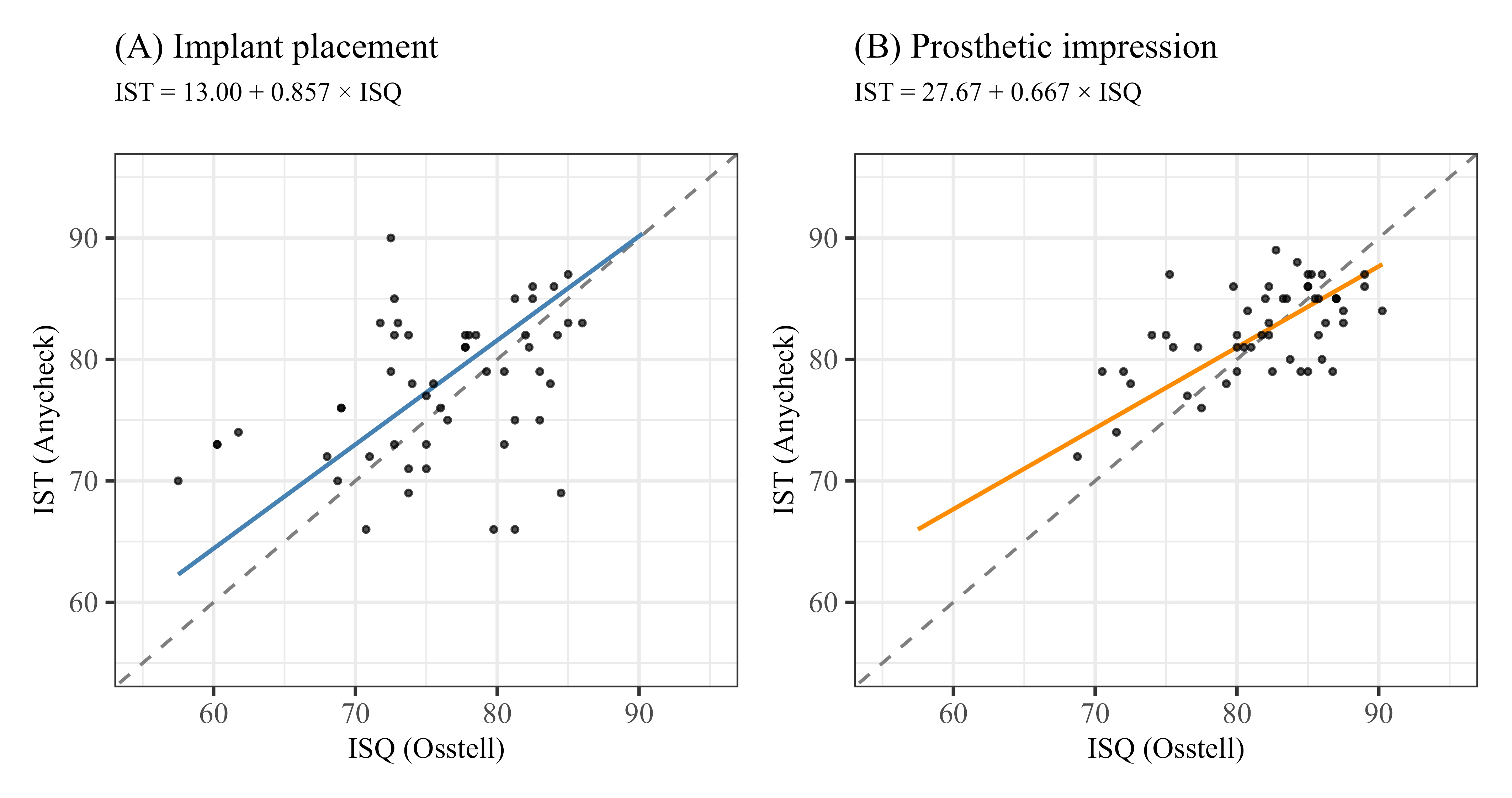
Fig. 1.
Scatter plots of ISQ versus IST measurements at two clinical time points with regression lines (dashed line, line of identity). (A) Implant placement, (B) prosthetic impression. Regression lines were fitted using Passing-Bablok regression, and the correlation coefficient r and p value in each panel were obtained from Pearson correlation analysis.
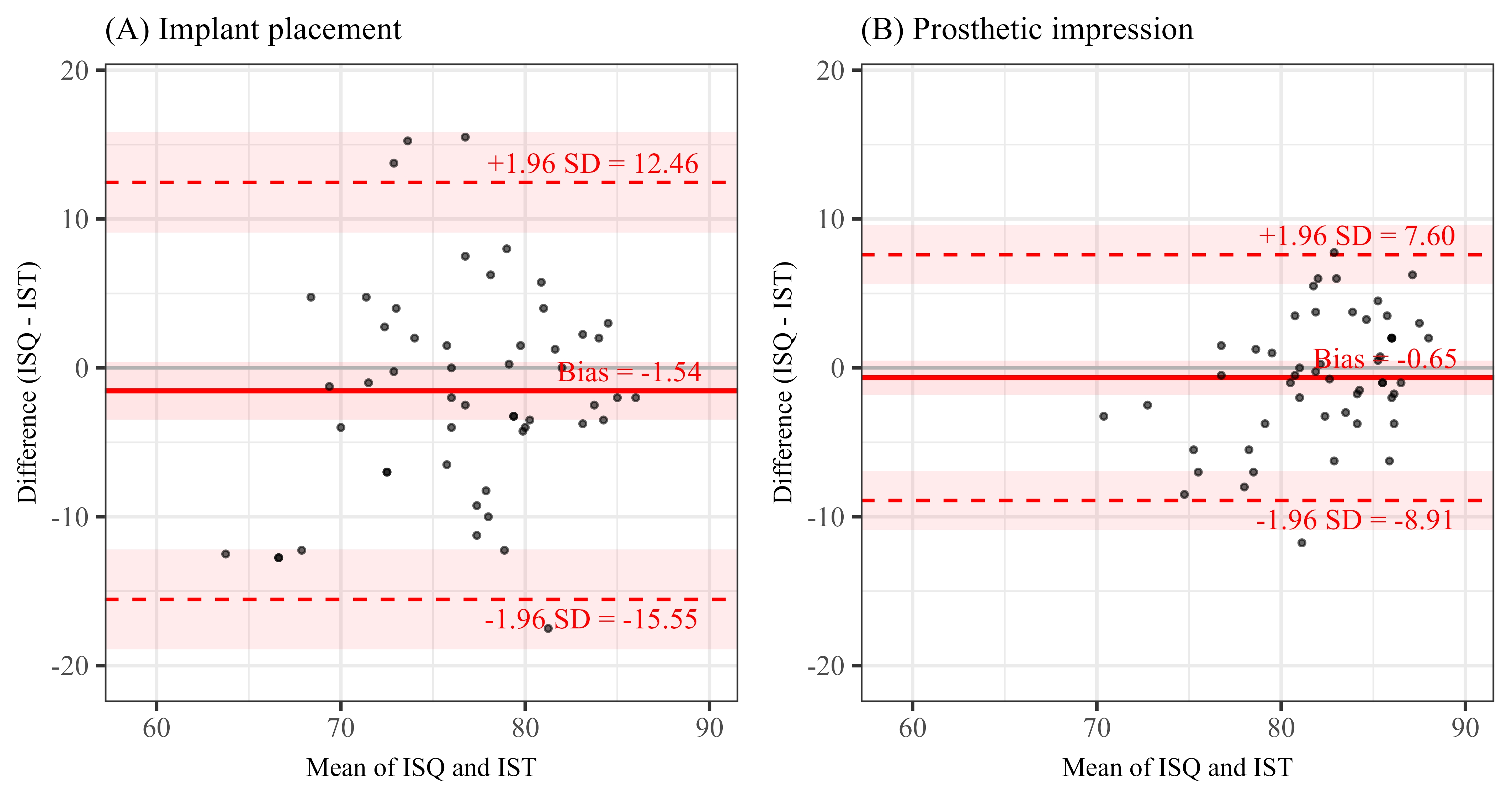
Figure 2 shows Bland–Altman plots comparing raw ISQ and IST values. On the original measurement scales, the mean bias between ISQ and IST was ‒1.54 at implant placement and ‒0.65 at prosthetic impression, neither of which was statistically significant (p = .126 and p = .268, respectively). The 95% LoA narrowed from implant placement to prosthetic impression, indicating greater agreement between the two devices as osseointegration progressed.
At implant placement, 25 implants were classified as stable by both devices and 11 as unstable by both devices; six were classified as stable only by ISQ and 10 only by IST. The percent agreement was 69.2% (95% CI: 55.7–80.1), with a Cohen’s κ of 0.341 (95% CI: 0.074–0.607). At prosthetic impression, 46 implants were classified as stable by both devices and two as unstable by both devices; none were classified as stable only by ISQ, whereas four were classified as stable only by IST. The percent agreement increased to 92.3% (95% CI: 81.8–97.0), and Cohen’s κ increased to 0.469 (95% CI: 0.019–0.920) (Table 2). Sensitivity analysis using a ≥70 cut-off showed agreement of 75.0% at implant placement and 98.1% at prosthetic impression (Table 3).
Table 2.
Categorical agreement between ISQ and IST classifications using a stability cut-off of ≥75 at implant placement and at prosthetic impression
| Agreement category | Implant placement | Prosthetic impression | |
| Concordant† | Both stable | 25 | 46 |
| Both unstable | 11 | 2 | |
| Discordant | ISQ stable only | 6 | 0 |
| IST stable only | 10 | 4 | |
| Percent agreement (95% CI) | 69.2% (55.7–80.1) | 92.3% (81.8–97.0) | |
| Cohen’s ꆆ (95% CI) | 0.341 (0.074–0.607) | 0.469 (0.019–0.920) | |
†Values for concordant and discordant categories are presented as the number of implants. ††Cohen’s κ values were interpreted following Landis and Koch (1977): 0.21–0.40 fair, 0.41–0.60 moderate. ISQ: Implant Stability Quotient (Osstell ISQ®); IST: Implant Stability Test value (AnyCheck®); CI: Confidence Interval.
Table 3.
Categorical agreement between ISQ and IST classifications using a stability cut-off of ≥70 at implant placement and at prosthetic impression (sensitivity analysis)
| Agreement category | Implant placement | Prosthetic impression | |
| Concordant† | Both stable | 39 | 51 |
| Both unstable | 0 | 0 | |
| Discordant | ISQ stable only | 5 | 0 |
| IST stable only | 8 | 1 | |
| Percent agreement (95% CI) | 75.0% (61.8–84.8) | 98.1% (89.9–99.7) | |
| Cohen’s ꆆ (95% CI) | ‒0.134 (‒0.397 to ‒0.128) | 0.000 (NE‡) | |
†Values for concordant and discordant categories are presented as the number of implants. ††Cohen’s κ values were interpreted following Landis and Koch (1977): 0.21–0.40 fair, 0.41–0.60 moderate. ISQ: Implant Stability Quotient (Osstell ISQ®); IST: Implant Stability Test value (AnyCheck®); CI: Confidence Interval.
Ⅳ. Discussion
The critical determinant of implant surgery success is achieving firm osseointegration and stability between the implant fixture and the surrounding bone. Historically, various methods, including push-out and push-through tests, removal torque analysis, and radiographic examinations, have been proposed to evaluate implant stability; however, most are either destructive or insufficiently accurate, limiting their utility for longitudinal assessment.18 To address these limitations, noninvasive approaches were developed, including the Periotest® system, which is based on DCA, and the Osstell® system, which uses RFA.3,19
Periotest® enables rapid measurements without consumables; however, its operator dependence limits reproducibility and precision,20,21,22 and direct percussion may induce micromovement in implants with low primary stability, potentially compromising osseointegration. Osstell ISQ®, which is currently widely accepted as the clinical standard, provides objective and highly reliable measurements using a noncontact measurement mechanism. Nevertheless, it requires removal of the healing abutment and attachment of a dedicated SmartPeg® at each measurement session, which can be clinically inconvenient.11 This is particularly relevant for immediate implant placement following tooth extraction using a flapless technique, in which repeated removal and replacement of components may introduce a significant confounding factor. If the fixture has not achieved sufficient primary stability during the early healing phase, the rotational torque generated by repeatedly screwing and unscrewing these components could potentially induce microtrauma at the bone–implant interface.4,23
In the present study, Cohen’s d was 1.09 for ISQ and 0.75 for IST, indicating large effect sizes for both measurements. Both ISQ and IST increased significantly from initial implant placement to prosthetic impression taking (Table 1), demonstrating that both parameters reliably reflected the progressive improvement in implant stability over time.24 Furthermore, the narrower distribution, indicated by smaller SDs, at the prosthetic impression stage than at initial placement suggests that implant stability converged toward a uniformly high level as osseointegration progressed.
Regression analysis evaluating the relationship between the two measurements demonstrated a positive linear association at both time points (Fig. 1). The most notable difference between the time points was the distribution of data points around the regression line. At initial placement, the data points were relatively widely scattered, whereas at the prosthetic impression stage, they were more tightly clustered within a narrower range. This pattern indicates reduced residual variability between the two measurements as osseointegration progressed, consistent with the narrower LoA observed in the Bland–Altman analysis. The change in the regression coefficient (from 0.857 to 0.667) should be interpreted in the context of the restricted stability range (approximately 70–90) observed at the prosthetic impression stage. The SD of IST decreased by approximately 38% (from 6.0 to 3.7), compared with a 24% reduction in ISQ (from 6.8 to 5.2). This asymmetric reduction in variability indicates that IST converged more rapidly toward the stable range than ISQ during osseointegration, which may account for the flatter regression slope. Accordingly, the decrease in the regression coefficient should not be interpreted as evidence of disagreement between the devices; rather, it reflects the convergence of both measurements toward their respective stable ranges as implant stability increased. The wider distribution of measurements at placement was likely attributable to interindividual variability in anatomical conditions, bone density, and residual bone volume within the extraction socket.25
The Bland–Altman analysis assessing clinical agreement between the two measurements (Fig. 2) showed mean biases of ‒1.54 at initial placement and ‒0.65 at the prosthetic impression stage, both of which were close to zero. However, a mean bias close to zero does not necessarily indicate that the two measurements were consistently similar; the 95% LoA must also be considered to assess the potential magnitude of discrepancy at the individual implant level.26 At implant placement, the width of the 95% LoA indicated a potential discrepancy of approximately 28 units, which narrowed to approximately 16 units at the prosthetic impression stage. These findings indicate that as osseointegration progressed and implant stability increased, the differences between the two measurements diminished, resulting in greater clinical agreement.
Given the clinical context of immediate implant placement following tooth extraction, a conservative stability threshold of ≥75 was adopted.14,15,16 Categorical agreement analysis (Table 2) showed that the percentage agreement between the two devices was 69.2% at implant placement and increased substantially to 92.3% at the prosthetic impression stage. This increase indicates improved agreement in stability classification between the devices. The relatively lower categorical agreement at implant placement (69.2%) was likely attributable to substantial inter-implant variability in primary stability arising from patient-specific differences in bone density, bone quality, and extraction socket anatomy.25 Furthermore, because the two devices use fundamentally different measurement principles—RFA for ISQ and DCA for IST—they may exhibit different sensitivities during the early postsurgical period, when primary stability is relatively low and variability is high.11 The finding that more implants were classified as stable by IST alone (n = 10) than by ISQ alone (n = 6) further suggests that the two devices may have different sensitivity profiles during the early healing phase. The concurrent use of both measurement modalities may therefore represent a more prudent clinical approach at this stage. In contrast, the high agreement observed at the prosthetic impression stage indicates that once osseointegration has progressed sufficiently, both devices provide highly concordant stability classifications, consistent with the Bland–Altman findings.27
In summary, ISQ and IST values changed in the same direction over time, and the discrepancy between the two measurements progressively decreased as osseointegration advanced. At the prosthetic impression stage, the two measurements converged more closely, suggesting that similar clinical judgments may be made regardless of the device used. Nevertheless, because the LoA at this stage still spanned approximately ±10 units, the two measurements should not be considered fully interchangeable. Therefore, longitudinal follow-up of an individual implant should ideally be performed using the same measurement instrument throughout the observation period.28
The present study has several limitations. First, the relatively small sample size (52 implants) resulted in wide 95% CIs for the agreement indices, including Cohen’s κ. Notably, at the prosthetic impression stage, only two implants were classified as unstable, resulting in a wide 95% CI (0.019–0.920) for the κ value (0.469). Future studies with larger sample sizes are needed to improve the precision of these estimates.29 Second, the two devices differ in their measurement procedures. Osstell ISQ® averages measurements obtained from four directions (mesial, buccal, distal, and lingual), whereas AnyCheck® measures stability only from the buccal aspect. Although both devices were used according to their respective manufacturer protocols, the potential effect of this methodological difference on agreement cannot be excluded. Third, direct biomechanical or histological validation methods, such as removal torque analysis or histologic evaluation, were not performed. Therefore, the extent to which either device reflects true osseointegration could not be directly established. Future studies incorporating an independent reference standard are warranted to address this limitation. Fourth, the study did not perform stratified analyses according to implant- and site-related variables, including implant diameter and length, insertion site, and the healing interval before prosthetic impression. In addition, insertion torque and bone quality, which are objective indicators of primary stability, were not consistently documented in the retrospective records and therefore could not be included in the analysis. Given the retrospective, single-center design and limited sample size, subgroup analyses would have had insufficient statistical power. Because these factors may influence the agreement between the two devices, large-scale prospective studies with standardized conditions are warranted to clarify their effects. Fifth, although comparison of the temporal changes in ISQ and IST from implant placement to prosthetic impression would more closely address the longitudinal aspect of this study, the two-time-point retrospective design precluded trajectory-based analysis. Future prospective studies with multiple measurement time points are needed to directly compare the temporal dynamics of the two devices.
Despite these limitations, the findings demonstrate that, even in challenging clinical scenarios such as immediate molar implant placement, in which primary stability is critical, both measurement modalities showed a consistent increase from implant placement to prosthetic restoration.27 Although Osstell ISQ® may remain the preferred reference method in research settings, AnyCheck® offers practical advantages for routine clinical use, including shorter measurement time and reduced risk of soft-tissue contact. These characteristics may allow clinicians to select the device that best suits their clinical requirements.
Ⅴ. Conclusion
Implant stability increased over time as measured by both Osstell ISQ® and AnyCheck®, with agreement between the devices improving as osseointegration progressed. Once osseointegration was established, the two devices provided clinically comparable measurements, although they were not fully interchangeable. Either device may therefore be used to support stability assessment at this stage. However, consistent use of the same device is advisable when monitoring an individual implant longitudinally.