


Ⅰ. Introduction
Post-extraction alterations of hard and soft tissues may result in labial soft tissue collapse, changes in the gingival margin, papilla deficiency, and open gingival embrasures. These changes are particularly critical in the maxillary anterior region, where the labial bone is often thin and esthetic demands are high.
Immediate implant placement may reduce dimensional alterations but cannot fully prevent post-extraction ridge remodeling.1,2,3 Therefore, customized healing abutments or provisional prostheses are often used to support the peri-implant soft tissues and establish an appropriate emergence profile.4,5,6 In tooth-supported fixed dental prostheses, an ovate pontic delivered immediately after extraction can also support the extraction socket and guide soft tissue healing at the pontic site.7,8,9 Although one site in this report was restored with an implant-supported prosthesis and the other with a tooth-supported prosthesis, both approaches share a common principle: a provisional prosthetic component placed immediately after extraction, before soft tissue collapse occurs, can serve as a scaffold to support the post-extraction soft tissues and facilitate transfer of the established contour to the definitive prosthesis. This report describes two cases involving anterior prostheses in which immediate provisional prosthetic components were used to maintain post-extraction soft tissue contours and transfer them to the definitive prostheses.
Ⅱ. Case Reports
1. Case 1
A 59-year-old man presented with a fractured crown of the maxillary left central incisor. During endodontic treatment, a crack line extending toward the root was identified, and extraction with immediate implant placement was planned (Fig. 1A to 1C). The gingival margin of the maxillary left central incisor was confirmed to be at the same level as that of the maxillary right central incisor. Preoperative cone-beam computed tomography confirmed that the site was suitable for immediate implant placement. A surgical guide was fabricated to position the implant approximately 4 mm apical to the gingival margin to achieve a favorable emergence profile (Fig. 1D).5
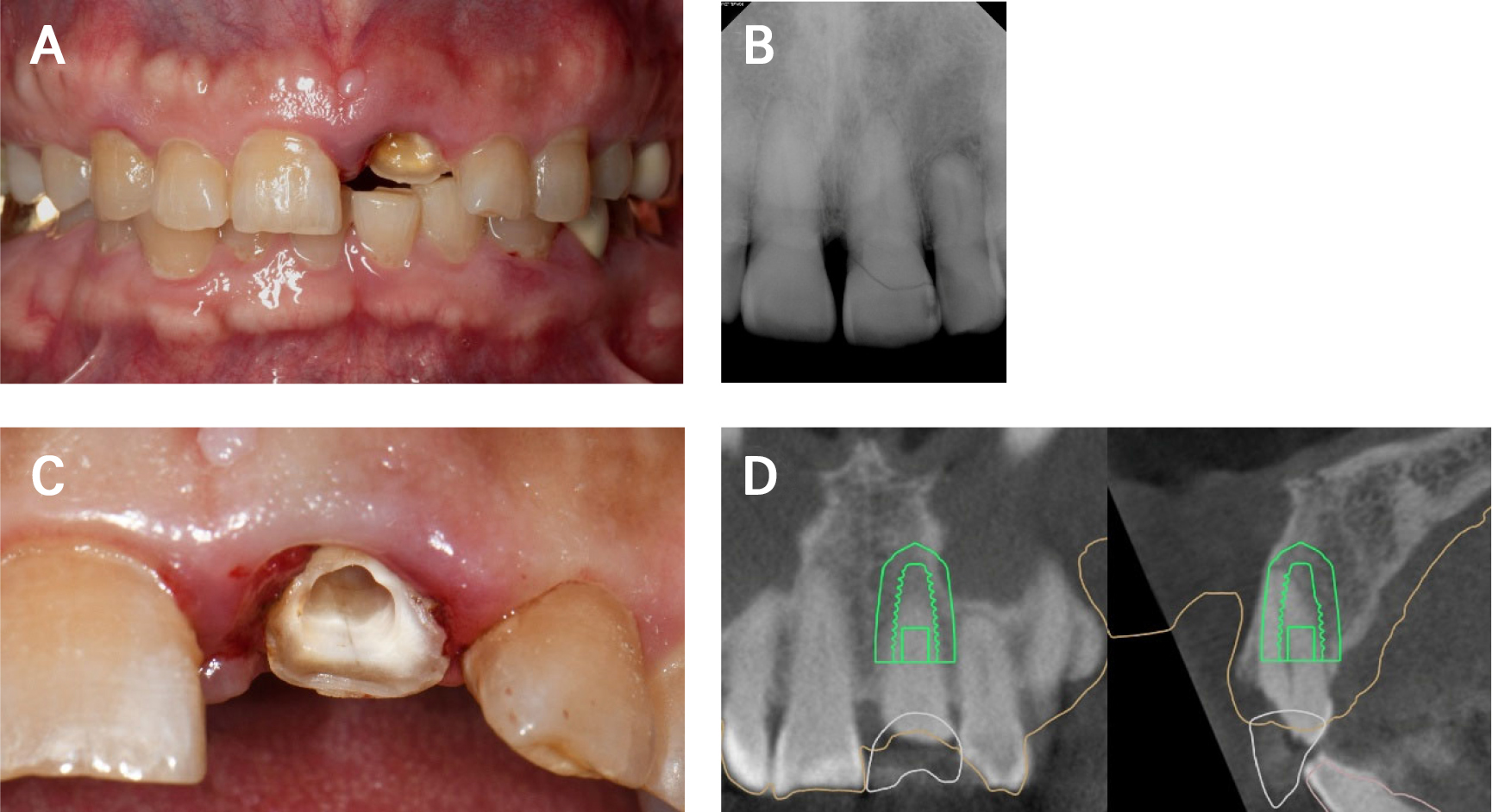
Fig. 1.
Initial examination and treatment planning for Case 1. (A) Initial intraoral photograph showing the fractured maxillary left central incisor, (B) Preoperative radiographic evaluation, (C) Identification of the crack line extending toward the root, (D) Treatment planning for immediate implant placement using a surgical guide.
Following atraumatic extraction, a 4.1 × 10-mm implant (BlueDiamond; MegaGen Implant Co., Ltd., Daegu, Korea) was placed using the surgical guide. The gap between the implant and labial bone was grafted with xenograft material (Bio-Oss; Geistlich Pharma, Wolhusen, Switzerland) (Fig. 2A and 2B). A temporary abutment was connected, and a customized healing abutment was fabricated chairside using flowable resin and delivered (Fig. 2C to 2E).10 The subcritical contour was designed to be concave to minimize pressure and provide adequate space for soft tissue healing, whereas the critical contour was slightly under-contoured to avoid premature compression of the marginal soft tissue.4,6The subcritical contour was not adjusted during the initial 3-month healing period to facilitate stable osseointegration.
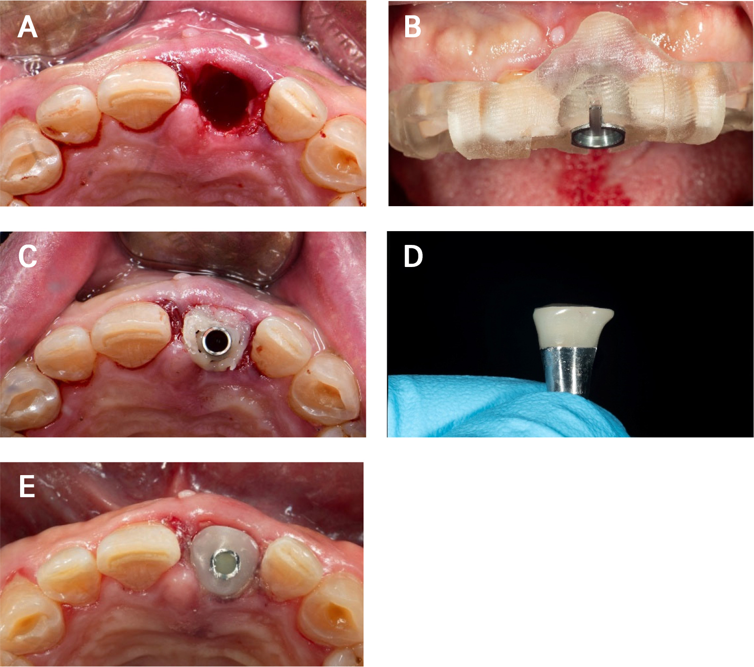
Fig. 2.
Surgical procedure and fabrication of the customized healing abutment for Case 1. (A) Atraumatic extraction of the maxillary left central incisor, (B) Implant placement using a surgical guide, (C) Chairside fabrication of the customized healing abutment using flowable resin, (D) Fabricated customized healing abutment, (E) Delivery of the customized healing abutment.
After 3 months, the peri-implant soft tissue contour was clinically confirmed to be stable. Because the customized healing abutment had been designed with a passive, concave subcritical contour, it remained undisturbed throughout the 3-month osseointegration period without further adjustment. The emergence profile was subsequently developed during the provisional phase to avoid compression of the peri-implant soft tissue during osseointegration. A customized impression coping was fabricated to reproduce the established contour on the working cast, and a screw-retained provisional prosthesis was delivered. Resin was then added to the critical contour, which had been slightly under-contoured on the customized healing abutment, to further develop the emergence profile and support the labial gingival margin (Fig. 3). For fabrication of the definitive prosthesis, intraoral scans of the provisional prosthesis in place, the provisional prosthesis itself, and the scan body were superimposed to transfer the subgingival contour to the definitive prosthesis (Fig. 4A to 4D).11 A screw-retained zirconia implant prosthesis was subsequently delivered (Fig. 4E). One month after delivery of the definitive prosthesis, the pre-extraction and post-delivery intraoral scans were superimposed using Exocad software (Exocad GmbH, Darmstadt, Germany). The scans were aligned using stable references, including nonmobile teeth and adjacent hard tissue, and the labial soft tissue contours were visually compared. This supportive visual assessment demonstrated relatively well-maintained labial soft tissue contours (Fig. 4F).
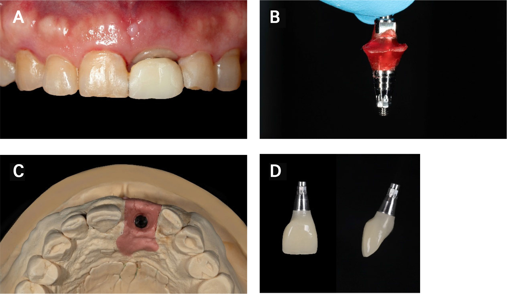
Fig. 3.
Transfer of the peri-implant soft tissue contour to the provisional prosthesis in Case 1. (A) Peri-implant soft tissue contour 3 months after surgery, (B) Customized impression coping, (C) Working cast reproducing the emergence profile established by the customized healing abutment, (D) Screw-retained implant provisional prosthesis.
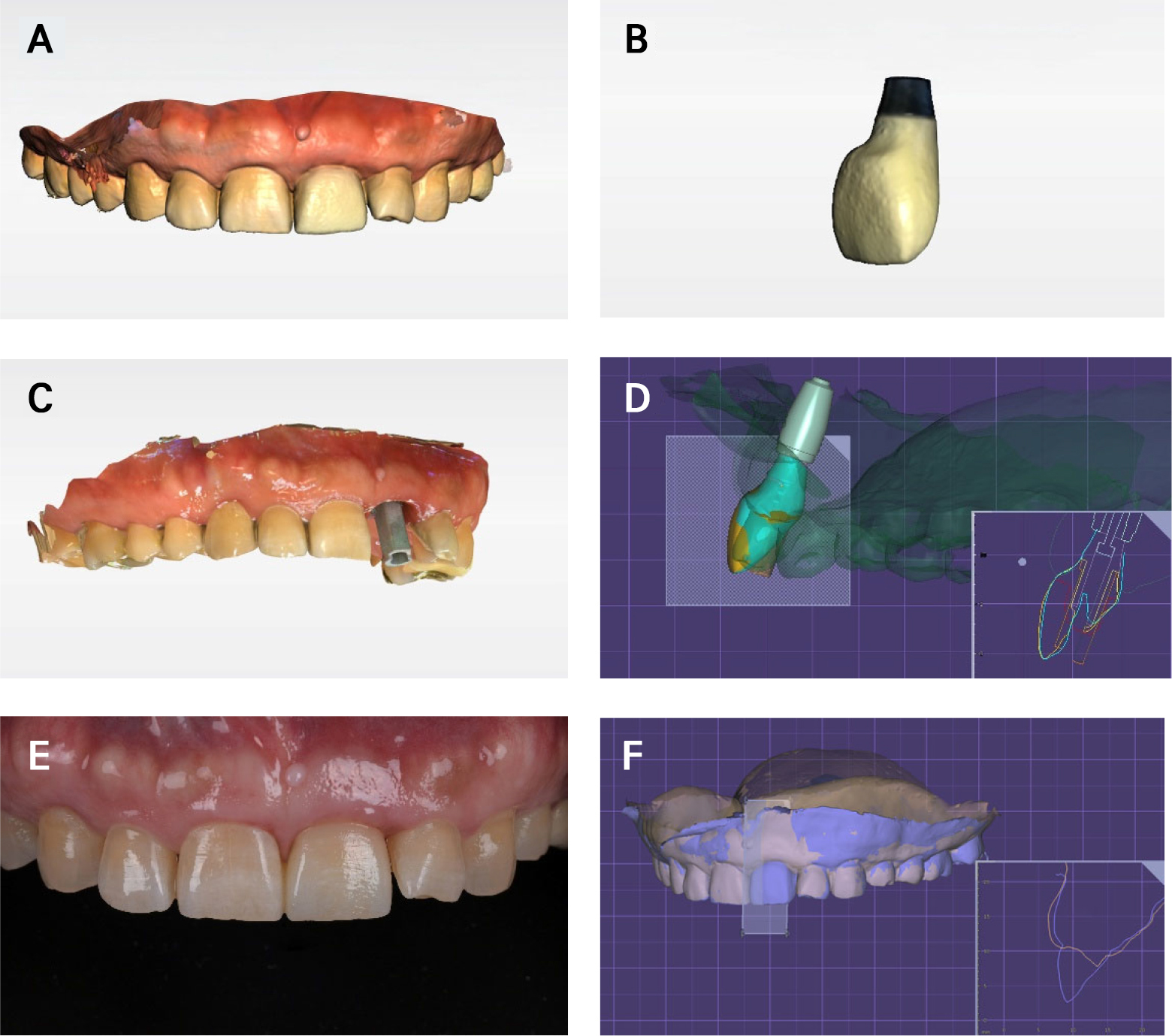
Fig. 4.
Digital transfer to the definitive prosthesis and visual assessment of the labial soft tissue contour in Case 1. (A) Intraoral scanning of the provisional prosthesis in place, (B) Scanning of the provisional prosthesis, (C) Digital impression of the implant with a scan body, (D) Definitive prosthesis design reproducing the subgingival contour of the provisional prosthesis, (E) Definitive screw-retained zirconia implant-supported fixed prosthesis 1 month after delivery, (F) Superimposition of pre-extraction and post-delivery scan data for visual comparison of the labial soft tissue contour.
2. Case 2
A 66-year-old man presented with a buccal fistula in the maxillary left lateral incisor region. Radiographic examination revealed a periapical lesion associated with the maxillary left lateral incisor. Removal of the existing splinted fixed prosthesis revealed deep palatal root caries affecting the maxillary right central incisor (Fig. 5A to 5D). Extraction of the maxillary right central incisor, replacement of the existing tooth-supported fixed dental prosthesis extending from the maxillary right lateral incisor to the maxillary left central incisor, endodontic retreatment, and placement of a crown prosthesis for the maxillary left lateral incisor were planned.
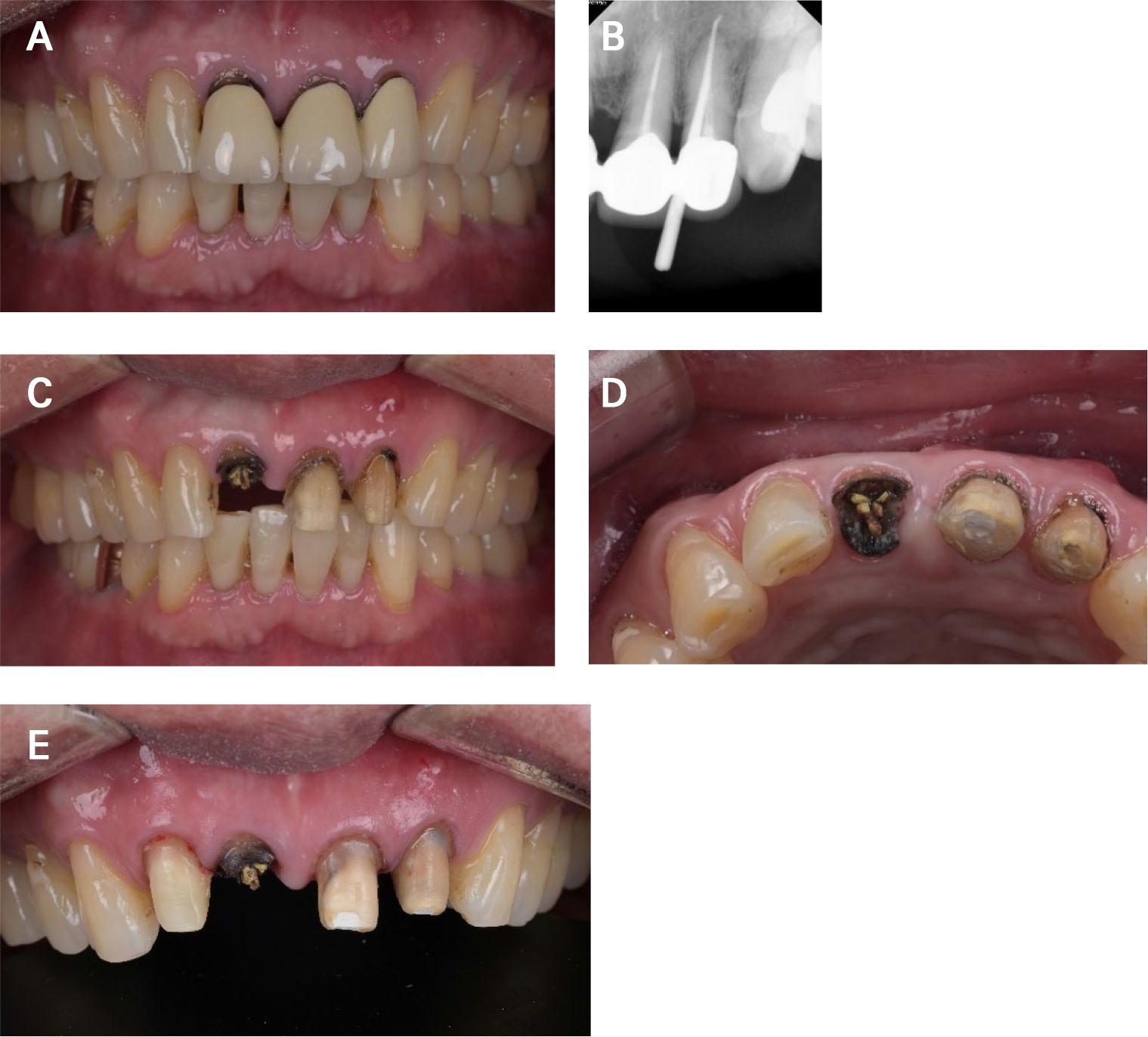
Fig. 5.
Initial examination and treatment planning for Case 2. (A) Initial intraoral photograph, (B) Preoperative periapical radiograph, (C) Intraoral photograph after removal of the existing fixed prosthesis, (D) Deep palatal root caries of the maxillary right central incisor, (E) Tooth preparation before extraction.
To facilitate immediate provisionalization, tooth preparation and intraoral scanning were performed before extraction (Fig. 5E). A provisional fixed dental prosthesis with an ovate pontic extending approximately 3 mm into the extraction socket was digitally designed and milled from polymethyl methacrylate (Fig. 6A to 6C).12,13 Following atraumatic extraction, alveolar ridge preservation was performed using 0.25 g of Bio-Oss (Geistlich Pharma) and a 13 × 25-mm Bio-Gide collagen membrane (Geistlich Pharma). The provisional prosthesis was then delivered immediately (Fig. 6D and 6E).9
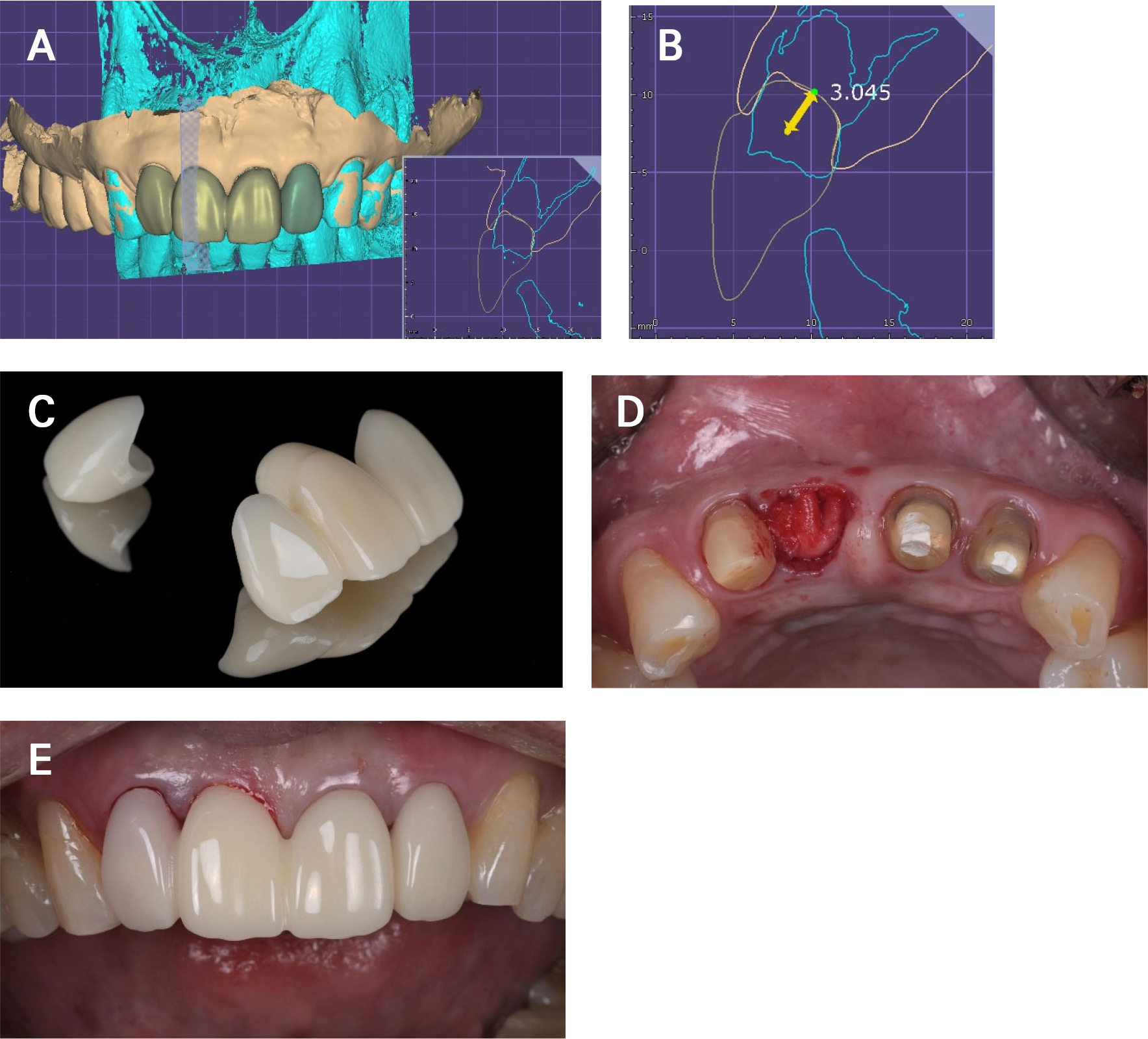
Fig. 6.
Immediate provisional fixed dental prosthesis with an ovate pontic in Case 2. (A) Design of the provisional prosthesis, (B) Ovate pontic extending approximately 3 mm into the extraction socket, (C) Milled polymethyl methacrylate provisional prosthesis, (D) Alveolar ridge preservation using xenograft material and a resorbable collagen membrane, (E) Delivery of the provisional prosthesis.
After 4 weeks, gingival asymmetry and insufficient papilla formation were observed. The provisional prosthesis was modified by adding resin to the subgingival area to enhance the labial gingival contour. The distal surface of the pontic was reduced, and resin was added mesially to shift the pontic in a mesial direction (Fig. 7). For fabrication of the definitive prosthesis, scans of the provisional prosthesis in place, the abutment teeth, and the provisional prosthesis were superimposed to transfer the pontic contour to the definitive prosthesis (Fig. 8A to 8D).11 A zirconia fixed dental prosthesis and a crown prosthesis were subsequently delivered (Fig. 8E). One month after delivery of the definitive prostheses, the pre-extraction and post-delivery intraoral scans were superimposed using Exocad software. The scans were aligned using stable references, including nonmobile teeth and adjacent hard tissue, and the labial soft tissue contour around the pontic site was visually compared. This supportive visual assessment demonstrated a relatively well-maintained soft tissue contour (Fig. 8F).
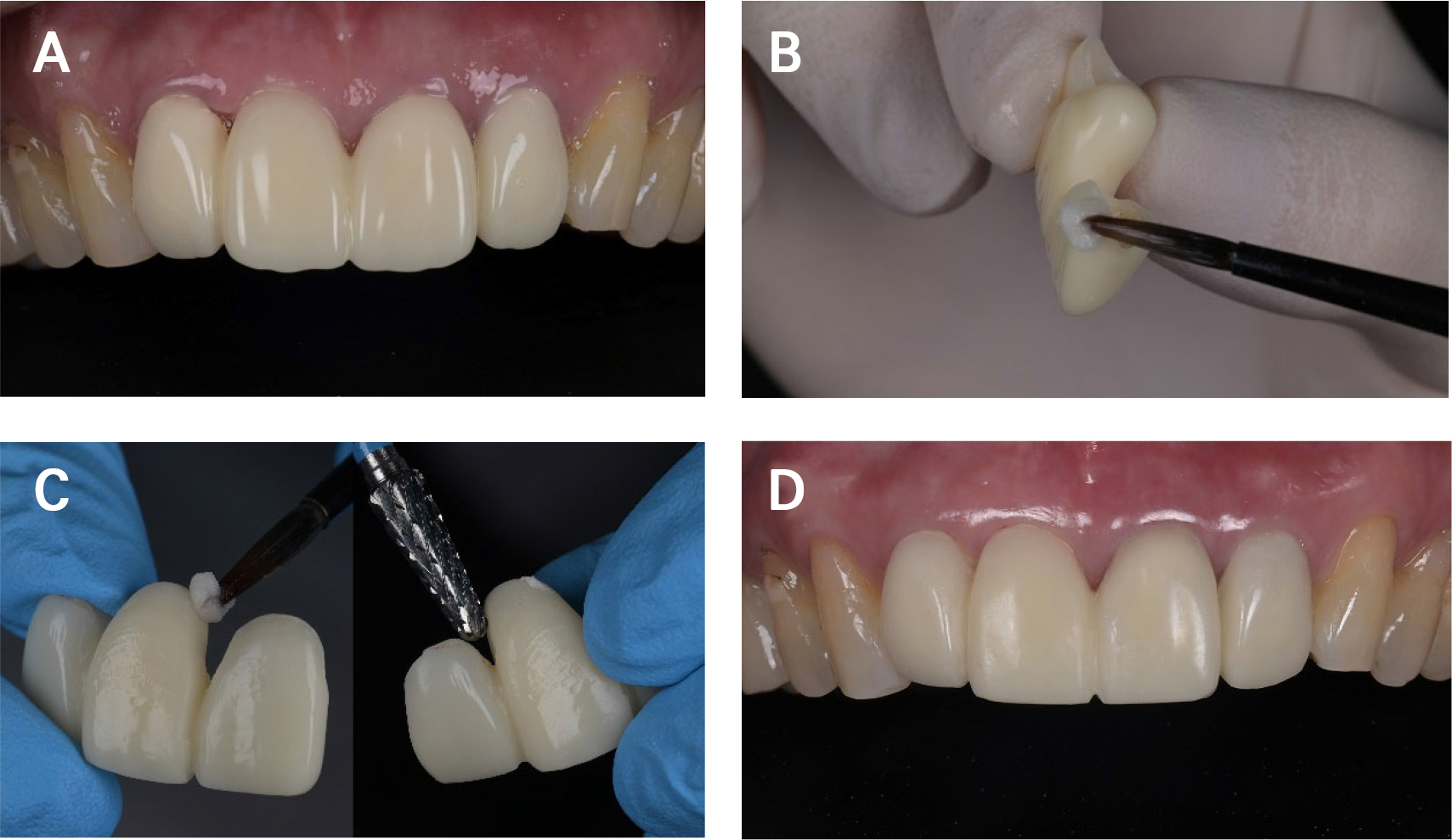
Fig. 7.
Modification of the provisional prosthesis for gingival contouring in Case 2. (A) Gingival contour 4 weeks after extraction, showing gingival asymmetry and insufficient interdental papilla formation, (B) Addition of resin to the subgingival area to enhance the labial gingival contour, (C) Mesial repositioning of the ovate pontic by reducing the distal area and adding resin to the mesial area, (D) Improved gingival contour and interdental papilla formation after modification.
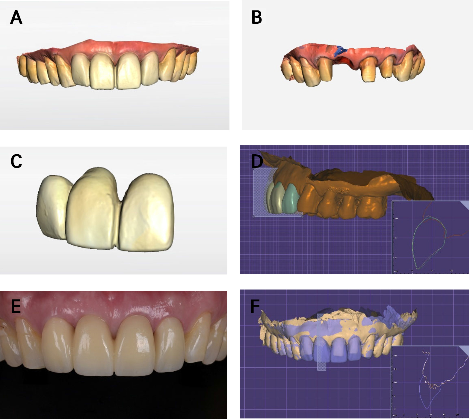
Fig. 8.
Digital transfer to the definitive prosthesis, delivery, and visual assessment of the labial soft tissue contour in Case 2. (A) Intraoral scanning of the provisional prosthesis in place, (B) Scanning of the abutment teeth, (C) Scanning of the provisional prosthesis, (D) Definitive prosthesis design reproducing the subgingival contour and pontic form of the provisional prosthesis, (E) Intraoral appearance 1 month after delivery of the zirconia fixed dental prosthesis and single crown prosthesis, (F) Superimposition of pre-extraction and post-delivery scan data for visual comparison of the labial soft tissue contour.
Ⅲ. Discussion
The two cases demonstrate the use of immediate provisional prosthetic components as scaffolds for maintaining post-extraction soft tissue contours in anterior esthetic restorations. Although the prosthetic designs differed, both approaches provided early soft tissue support and enabled transfer of the provisional prosthesis contour to the definitive restoration. Case 1 involved peri-implant soft tissue management following immediate implant placement, whereas Case 2 involved pontic-site development following extraction and alveolar ridge preservation. The cases were considered together because they share a common strategy: immediate placement of a provisional prosthetic component to support the extraction-site soft tissues before collapse, followed by transfer of the developed contour to the definitive prosthesis.
In Case 1, the customized healing abutment was fabricated after implant placement, allowing its contour to be adjusted according to the actual implant position and socket morphology. A concave subcritical contour and a slightly under-contoured critical contour were used to minimize soft tissue compression during healing.4,6 The established peri-implant contour was subsequently reproduced using a customized impression coping and superimposition of the provisional prosthesis scan data.
In Case 2, an ovate pontic was delivered immediately after extraction and alveolar ridge preservation. Previous reports suggest that immediate placement of an ovate pontic, particularly when combined with ridge preservation, may attenuate dimensional ridge changes and help maintain esthetic soft tissue contours. When placed immediately into the extraction socket, the ovate pontic supports the grafted site and surrounding soft tissues during healing. Thus, the pontic and alveolar ridge preservation procedure may have complementary effects, with the pontic limiting early soft tissue collapse while the graft helps maintain the underlying ridge dimensions.8,9,14 Because the soft tissue contour changed during healing, the provisional prosthesis was modified at 4 weeks to improve gingival symmetry and papillary form before fabrication of the definitive prosthesis.
This report has several limitations. First, only two cases were included, and the prosthetic approaches differed between the cases. Second, errors related to intraoral scanning, scan-data alignment, and soft tissue measurements cannot be excluded. Soft tissue changes were assessed primarily through visual evaluation of superimposed scans rather than validated quantitative measurements. Third, follow-up was limited to approximately 1 month after delivery of the definitive prosthesis; therefore, only early outcomes could be evaluated. In addition, bone grafting was performed in both cases and may have contributed to preservation of the underlying ridge dimensions and labial soft tissue contours. Thus, the observed contour maintenance cannot be attributed solely to the provisional prosthetic components, and the relative contributions of grafting and prosthetic soft tissue support cannot be determined from these case reports. Longer-term follow-up is necessary to evaluate the biological and mechanical stability of the outcomes. Nevertheless, these cases suggest that atraumatic extraction, ridge preservation when indicated, appropriate subgingival contour design, provisional modification, and accurate transfer of the provisional contour may collectively contribute to predictable anterior esthetic outcomes.
Ⅳ. Conclusion
Within the limitations of this report, immediate provisional prosthetic components were useful for supporting post-extraction soft tissue contours in anterior esthetic restorations. In the implant-supported prosthesis case, a customized healing abutment and provisional prosthesis supported and maintained the peri-implant soft tissue contour. In the tooth-supported fixed dental prosthesis case, an ovate pontic supported the extraction socket and guided pontic-site development. Digital transfer of the provisional prosthesis contour facilitated fabrication of the definitive zirconia prostheses while maintaining relatively stable labial soft tissue contours.