


Ⅰ. Introduction
Untreated odontogenic infections can progress beyond localized inflammation, leading to severe complications such as cutaneous fistula formation in the maxillofacial region. Similarly, inflammatory conditions associated with dental implants facilitate infection spread, allowing pathogens to penetrate the surrounding alveolar bone, breach the cortical plate, and extend into adjacent soft tissues, potentially resulting in a maxillofacial fascial space abscess.1,2,3 In severe cases, these infections can cause extensive facial swelling, necrosis of the overlying soft tissue, and eventually cutaneous fistula formation. Although oral and maxillofacial infections are commonly polymicrobial, they are typically associated with oral flora, such as Streptococcus, Staphylococcus, Pseudomonas, and other species. In contrast, Citrobacter spp. rarely cause oral infections.4,5,6
Citrobacter is a genus of gram-negative, facultatively aerobic bacteria commonly found in the gastrointestinal tract. It is recognized as an opportunistic pathogen primarily associated with nosocomial infections.7,8,9 This case report describes a rare implant-related maxillofacial infection associated with C. freundii and C. braakii, presenting as a submandibular space abscess with a cutaneous fistula in an elderly patient.
Ⅱ. Case Report
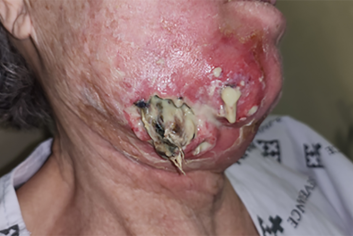
An 85-year-old woman was referred from the Department of Emergency Medicine to the Department of Oral and Maxillofacial Surgery at the Yonsei University Severance Hospital because of right submandibular swelling and skin necrosis (Fig. 1). Clinically, the patient exhibited a toxic appearance with extensive skin necrosis, redness, and purulent discharge from the overlying right mandible. The patient had trismus; however, no significant dyspnea or dysphagia was observed. The patient’s vital signs were stable. Her medical history was notable for hypertension, but was otherwise unremarkable. The patient had undergone placement of a right mandibular posterior dental implant at a local clinic 4 months earlier. Five days before the presentation, the healing abutment was tightened. Three days later, facial swelling developed, and extraoral incision and drainage (I & D) were performed at the same clinic, followed by presentation to the emergency room.
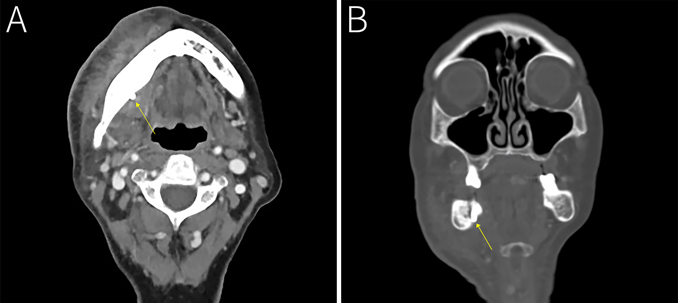
Laboratory tests performed in the emergency department revealed significantly elevated C-reactive protein levels and white blood cell counts (Table 1). Blood glucose levels and bone turnover markers were within normal limits. Contrast-enhanced computed tomography revealed a diffuse swelling and abscess formation in the right submandibular region. The infection extended from the implant site through the right lingual plate to the overlying cutaneous fistula (Fig. 2).
Table 1.
Laboratory test results upon arrival at the emergency room
The patient was admitted and treated with empirical intravenous antibiotics including cefoxitin (1 g t.i.d.) and metronidazole (500 mg t.i.d.). Wound care was provided and a purulent specimen was obtained for microbial culture. Aerobic, anaerobic, and fungal cultures were performed, and the results were reported on hospital day 5. Aerobic culture identified S. constellatus, C. braakii, and C. freundii, whereas no organisms were isolated from anaerobic or fungal cultures. Following consultation with the Department of Infectious Diseases, the antibiotic regimen was immediately adjusted to ampicillin/sulbactam (3 g q.i.d.) and ciprofloxacin (400 mg b.i.d.), based on antimicrobial susceptibility testing (Table 2).
Table 2.
Antimicrobial susceptibility testing of Citrobacter species
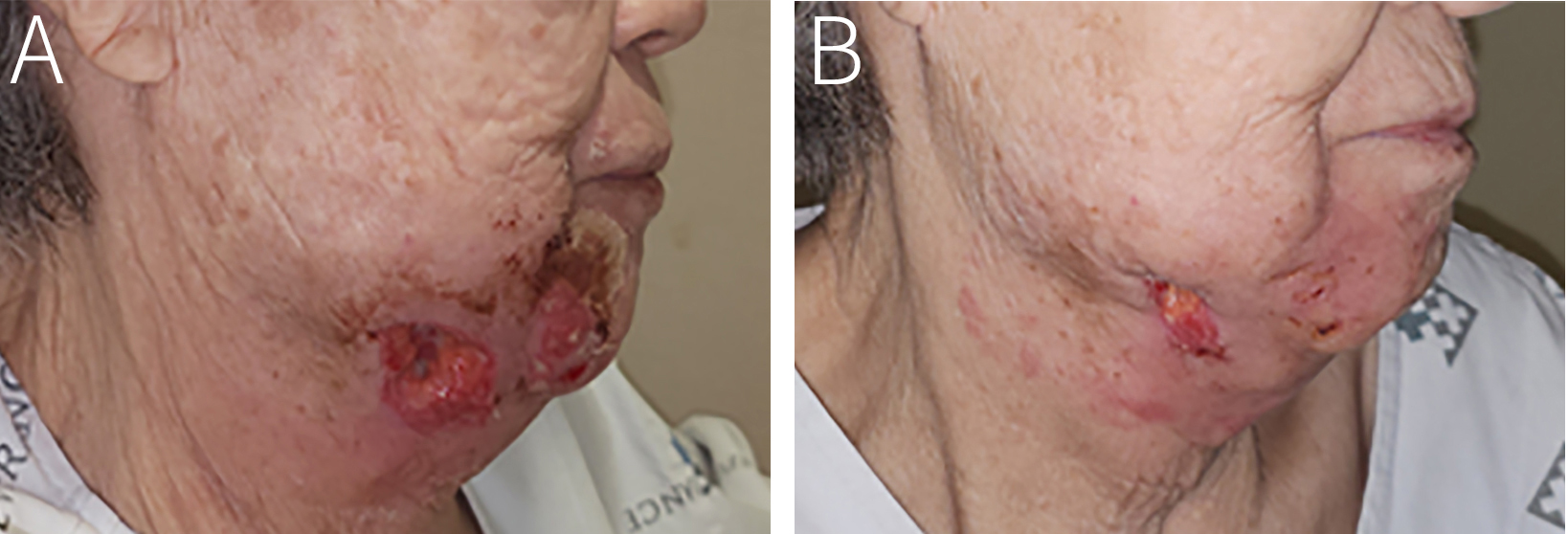
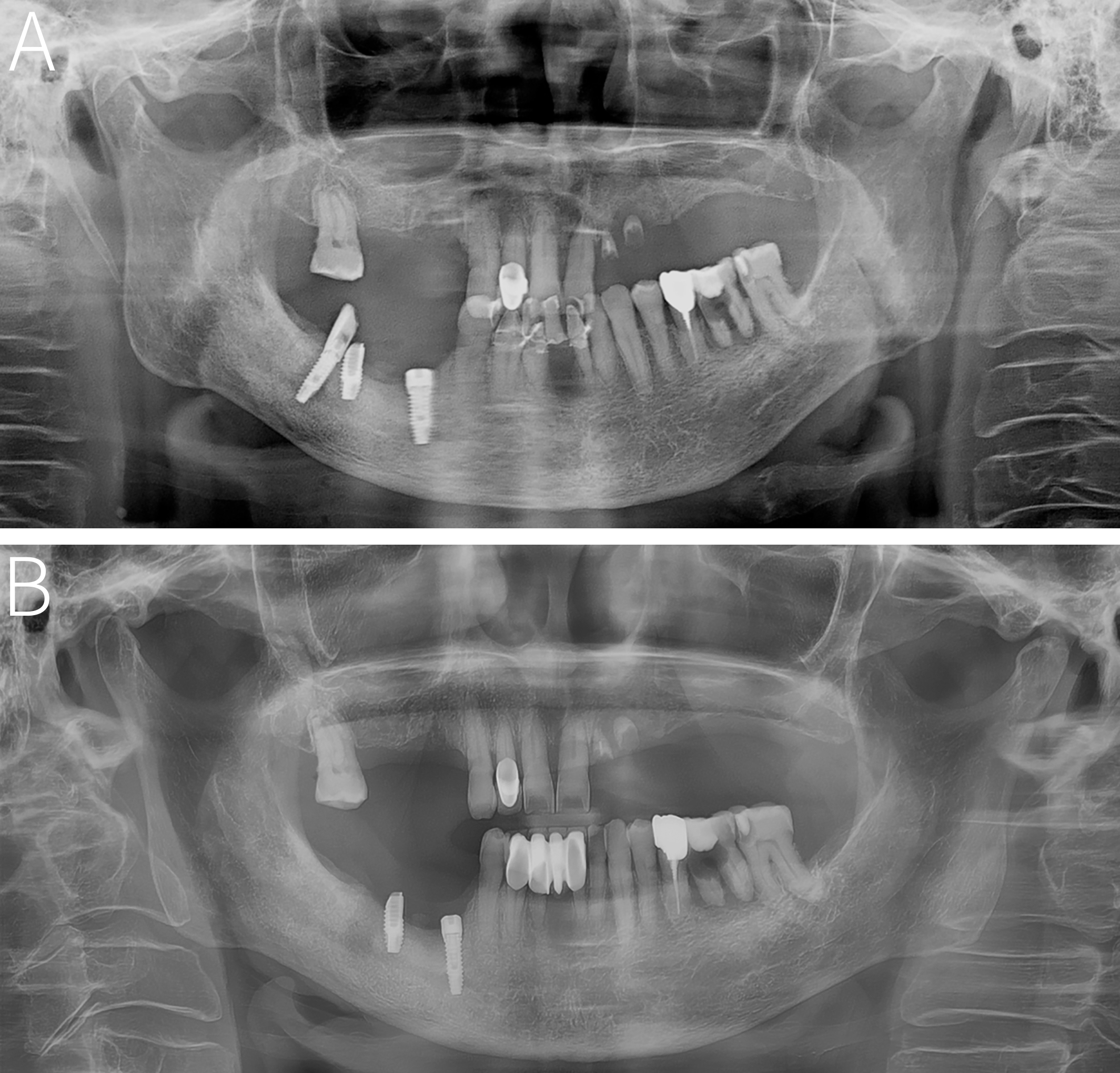
Following antibiotic adjustment, the patient improved considerably, with normalization of laboratory findings. The patient was discharged on hospital day 7 in good general condition (Fig. 3). Three days after discharge, the patient returned to the outpatient department where the dental implant was removed under local anesthesia. At that time, the infection further improved and most of the clinical symptoms subsided (Fig. 4). The patient continued antibiotic therapy with regular outpatient follow-ups and the wound healed without further complications.
Ⅲ. Discussion
Most maxillofacial infections are odontogenic in origin and commonly arise from periodontal disease, endodontic infections, or third molar-related pathology. Although dental implants are widely used for the rehabilitation of edentulous areas, they can be associated with infectious complications. Peri-implant infections are typically polymicrobial and involve inflammation of the tissues surrounding dental implants, which may be accompanied by progressive loss of supporting bone.10 In severe cases, infection may extend beyond the peri-implant tissues into adjacent maxillofacial fascial spaces, potentially resulting in serious complications. In this case, the dental implant extended through the lingual cortical plate. However, because immediate postoperative radiographs were unavailable, it was impossible to determine whether pre-existing implant malposition or biomechanical factors contributed to the lingual plate perforation and subsequent infection.
Odontogenic infections are typically polymicrobial, reflecting the diverse microbial flora of the oral cavity, and empirical antibiotic therapy is generally directed toward the predominant oral pathogens.11 In this case, microbial culture identified C. braakii and C. freundii in addition to S. constellatus. However, no organisms have been isolated from anaerobic or fungal cultures. However, negative anaerobic cultures may not exclude the involvement of anaerobic microorganisms because of the limitations in specimen collection and culture conditions. Antimicrobial susceptibility testing demonstrated resistance to ampicillin and cefoxitin, which were among the antibiotics initially selected for empirical treatment. Selection of appropriate antibiotics is particularly important when atypical or antimicrobial-resistant organisms are involved. Second-generation cephalosporins and metronidazole were initially selected empirically in our case; however, the regimen was subsequently adjusted to ampicillin/sulbactam and ciprofloxacin based on antimicrobial susceptibility testing. The patient recovered remarkably and was discharged on hospital day 7 with continued wound care and appropriate antibiotic therapy, without the need for additional surgical procedures, including further I & D or debridement.
Although Citrobacter spp. are exceptionally rare in odontogenic infections, their identification in this case highlights the importance of considering atypical organisms in severe implant-related infections.5, 6, 12Citrobacter spp. are predominantly associated with nosocomial infections and are rarely reported in the maxillofacial region.5,6,7,8, 11 Among the clinically recognized species, C. koseri has been reported in various healthcare-associated infections, whereas C. braakii and C. freundii were identified in our patient. A case of endophthalmitis following secondary intraocular lens implantation has been reported, with C. koseri identified as the causative microorganism.13 Similarly, Citrobacter spp. have rarely been reported in association with internal prosthetic implants. Kaufman et al. reported a case of C. koseri isolated from periprosthetic tissue 3 weeks after total hip arthroplasty, specifically from a hematoma in the femoral head region.14 Similarly, S. Craxford et al. documented a deep surgical site infection following hip hemiarthroplasty, in which Citrobacter spp. were identified in only 1 of 43 cases. The patient responded favorably to debridement, antibiotics, and implant retention.15
Furthermore, Citrobacter spp. have been implicated in infections involving fracture fixation implants. Ma et al. reported an implant-associated infection following surgery of a fracture and identified multiple bacterial species at the debridement site.16 Among the 41 bacterial species detected, C. braakii was less frequent, suggesting its potential involvement in implant-associated infection. Similarly, Yano et al. reported that sonication fluid culture following the removal of osteosynthesis materials was more effective for detecting microorganisms than tissue culture, which had the lowest detection rate.17 A previous study also reported Citrobacter spp. colonization on orthodontic mini-screws, suggesting a potential interaction with titanium surfaces.18, 19
However, considering that Citrobacter spp. are primarily found in the gastrointestinal tract and are more commonly implicated in urinary tract and nosocomial infections, their potential affinity for titanium-based dental implants and their role in peri-implant infections remain unclear. Future studies are warranted to clarify the potential interactions between Citrobacter spp. and dental implant materials and determine their clinical significance in implant-related infections.
Ⅳ. Conclusion
To the best of our knowledge, this is the first reported case of C. braakii and C. freundii infections in a patient with severe maxillofacial infections associated with dental implants. Considering the rarity of Citrobacter infections among odontogenic infections, the empirical selection of antibiotics may be challenging. In the present case, the isolation of Citrobacter spp. alongside S. constellatus suggested a polymicrobial infection, and antimicrobial susceptibility testing enabled the appropriate adjustment of antibiotic therapy. The patient achieved clinical resolution without additional surgical intervention, highlighting the importance of microbiological evaluation and susceptibility-guided treatment for severe infections.