I. 서론
II. 증례 보고
Ⅲ. 총괄 및 고찰
Ⅳ. 결론
I. 서론
임플란트 보철물은 유지 형태에 따라 크게 나사 유지형(screw-retained)과 시멘트 유지형(cement-retained)으로 나눌 수 있다1. 두가지 유지 형태는 상대적인 장단점이 있으며, 기술적 및 생물학적 합병증, 그리고 보철물의 기계적 안정성에 영향을 줄 수 있다2-5. 또한, 이러한 유지 형태는 임플란트의 가장 적절한 위치를 결정하기 위해 수술 전에 선택되어야 한다6. 하지만 아직까지 임상에서는 주로 술자의 선호도나 경험에 따라 임플란트 보철물의 유지 형태가 결정된다4.
나사 유지형 보철물은 retrievability가 우수하여 보철물의 수리 및 유지관리가 용이하고, 시멘트를 사용하지 않기 때문에 잔류 시멘트로 인한 임플란트 주위염, 부종, 궤양과 같은 합병증을 막을 수 있다3, 7. 실제로 이전의 전향적 임상시험에서 시멘트 유지형 보철물에 비해 양호한 임플란트 주위 연조직 반응이 관찰되었다고 보고하였다8. 반면, 나사 유지형 보철물의 단점으로는 나사 풀림 및 파절과 같은 기계적인 문제가 발생할 수 있으며, 교합면 나사 구멍으로 인해 비심미적이고 불안정한 교합 접촉을 초래한다2, 3, 9. 또한, 보철물의 수동적 적합(passive fit)을 위해서 고도의 정밀도가 필요하고, 시간이 경과함에 따라 수동적 적합이 상실되는 문제가 있다10, 11.
시멘트 유지형 보철물은 교합면 나사 구멍이 없어서 안정적인 교합 접촉과 심미성을 부여할 수 있고, 기공과정이 간단할 뿐만 아니라 비용적인 측면에서도 이점이 있다2, 12, 13. 또한, 시멘트층이 충격 흡수재의 역할과 더불어 제작과정에서의 오차를 보상하여 수동적 적합을 부여하기 때문에 보철물/임플란트/치조골 전체에 걸쳐 균일한 응력 전달에 유리하다9, 12. 그러나 시멘트 유지형 보철물은 보철물의 수리 및 유지관리를 위한 탈부착(retrievability)이 어렵고, 잔류 시멘트로 인한 생물학적 합병증과 더불어 유지력에 대한 조절이 어렵다는 단점이 있다13, 14.
이와 같은 문제점들을 극복하기 위해, 국내에서는 나사 유지형과 시멘트 유지형 보철물의 장점을 결합한 나사-시멘트 유지형 보철물(screw-and-cement-retained prosthesis; SCRP)이 소개되었다10. 하지만, 이러한 보철물 또한 나사 구멍이 교합면 내로 위치해야 하기 때문에 임플란트 식립 위치와 각도에 따라 모든 경우 사용할 수 없고, 장기간 사용에 따른 시멘트 파손(washout)이 발생할 수 있다10, 15.
최근에는 기존의 임플란트 보철물의 단점을 극복하고, 술자가 원할 시 손쉽게 보철물을 탈부착 할 수 있는 새로운 유지 형태의 임플란트 보철 시스템(EZ Crown system; EZC, Samwon DMP, Yangsan, Korea)이 소개되었다16. EZC는 지르코니아 볼(zirconia ball)과 니켈-티타늄 스프링(NiTi spring)으로 구성된 조립형 attachment와 이러한 볼이 위치하는 유지구(retention groove)가 형성된 abutment로 구성되어 있다(Fig. 1). 따라서, 보철물이 retention screw 없이 체결되기 때문에 나사 풀림 및 파절과 같은 기계적 합병증을 제거할 수 있고, 보철물의 합착 후 구강 외에서 잔류 시멘트의 제거가 가능하므로 임플란트 주위염 등의 생물학적 합병증을 막을 수 있다. 또한 보철물은 술자에 의해 언제든지 착탈이 가능하여 유지 관리가 용이하다. 본 증례에서는 EZC를 이용하여 고정성 보철물을 수복한 증례를 소개하고 그의 임상적 유효성에 대해 생각해 보고자 한다.
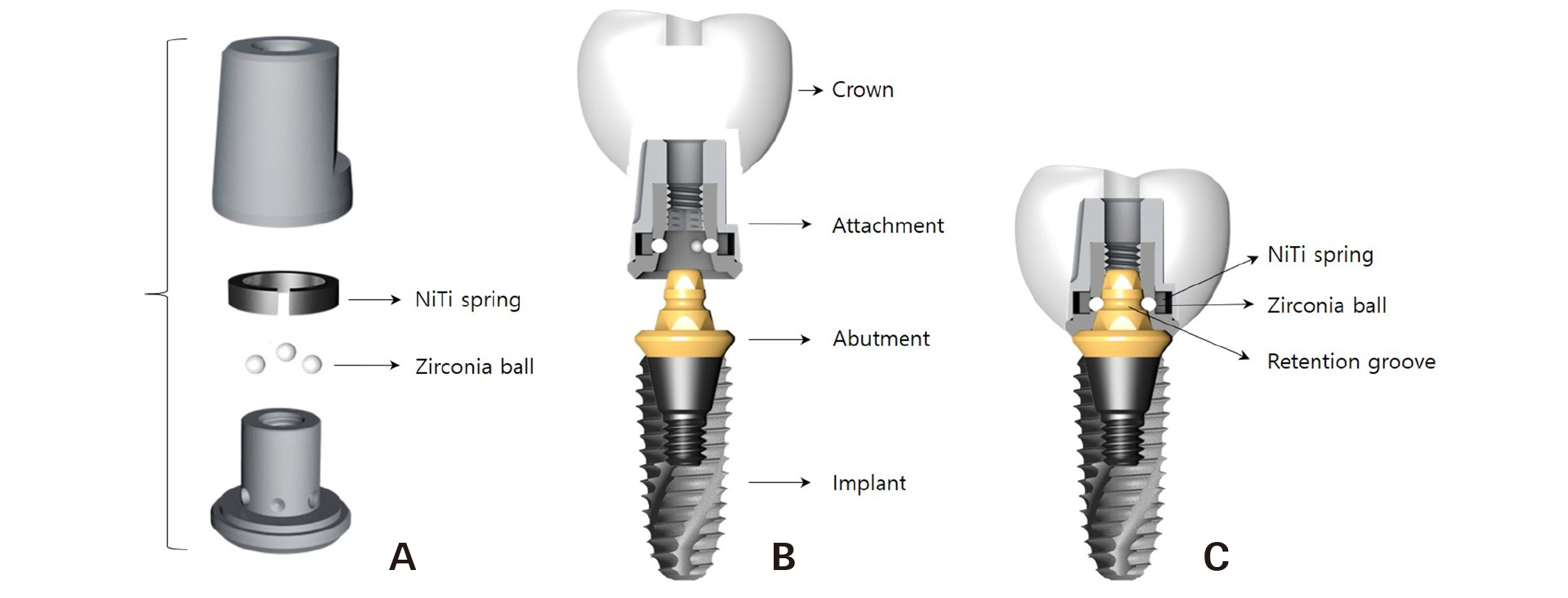
Fig. 1
(A) Sub-components of attachment, (B) Overall concept of the EZC, (C) Schematic representation of a final prosthesis fabricated with EZC.
Jae-Won Choi et al. : Implant-Supported Fixed Dental Prostheses with New Retention Type Using Zirconia Ball and Nickel-Titanium Spring. Implantology 2019
II. 증례 보고
본 증례는 56세 여자 환자로 기존의 보철물이 흔들리고 저작 시 아프다는 주소로 본원에 내원하였다. 치과적 병력으로 20년 전부터 #34=37 4-unit metal bridge를 착용하였으며, 특이할 만한 전신병력은 없었다. 지대치의 동요도와 방사선 검사 결과를 고려하여 #37을 발거하기로 하였다. 또한 인상 채득 및 진단용 납형을 제작하고 평가하여 무치악 부위에 두 개의 임플란트를 식립 후 고정성 임플란트 보철물을 제작하기로 최종 결정하였다. #37 발거 약 3개월 후 상하악 인상 채득하고 모형 제작하였다. 임플란트 식립부위의 잔존 치조골량과 정확한 식립위치를 평가하기 위해 Cone beam computed tomography (CBCT) 촬영하였다. 무피판 임플란트 수술(flapless implant surgery)을 계획하고, 제작한 모형과 CBCT 영상을 이용하여 surgical guide를 제작하였다.
Surgical guide를 이용하여 #35, 36 site에 직경 4.5 mm, 길이 10 mm, 직경 5.0 mm, 길이 7 mm 매몰형 (submerged) 임플란트(TS III, Osstem Implant Co, Busan, Korea)를 각각 식립하였다. 모두 35 Ncm 이상의 양호한 초기 고정력을 얻었으며 식립 후 치유 지대주(healing abutment)를 체결하였다(Figs. 2, 3).
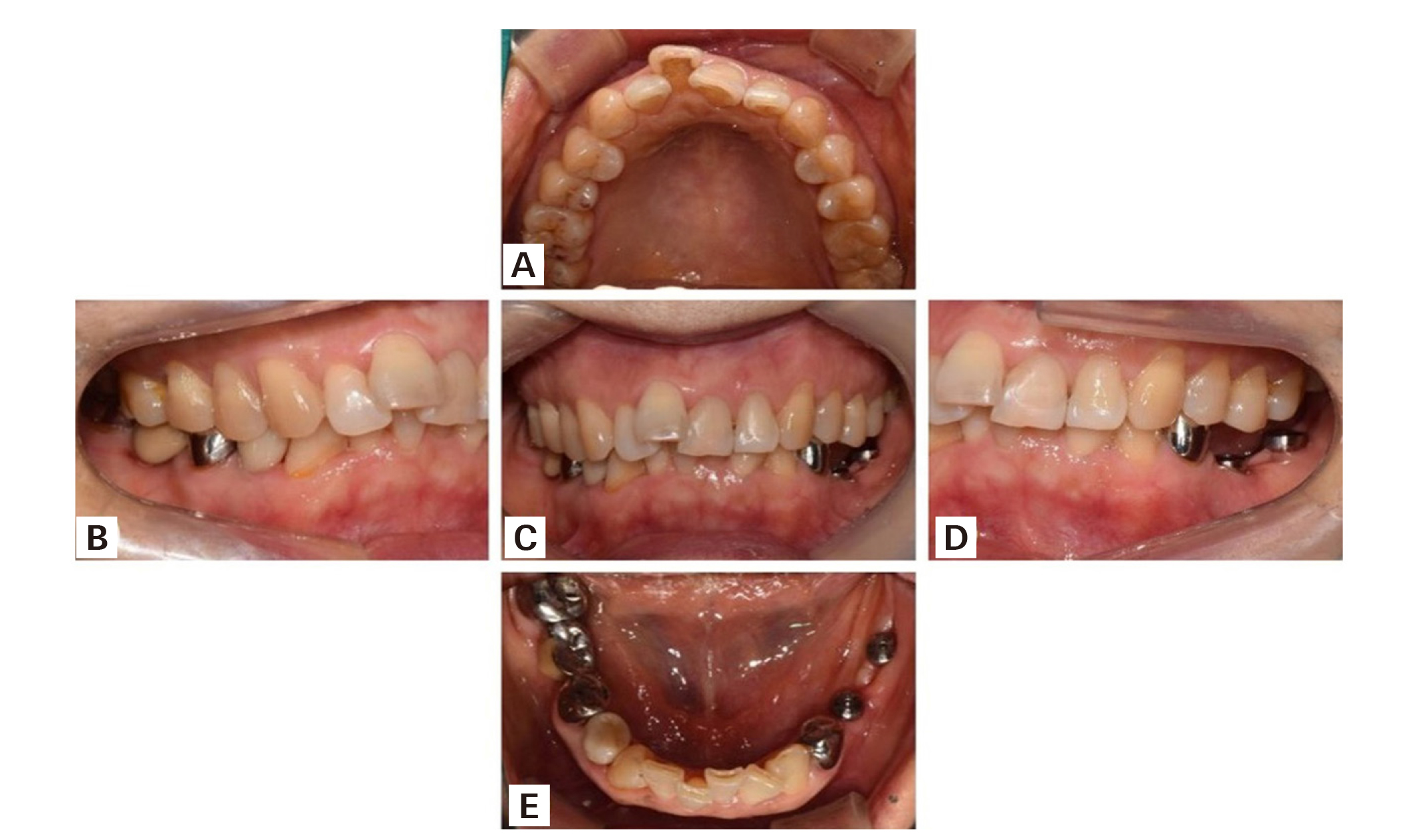
Fig. 2
AIntraoral view after implant installation. (A) Upper occlusal view, (B) Right lateral view, (C) Frontal view, (D) Left lateral view, (E) Lower occlusal view.
Jae-Won Choi et al. : Implant-Supported Fixed Dental Prostheses with New Retention Type Using Zirconia Ball and Nickel-Titanium Spring.
Implantology 2019
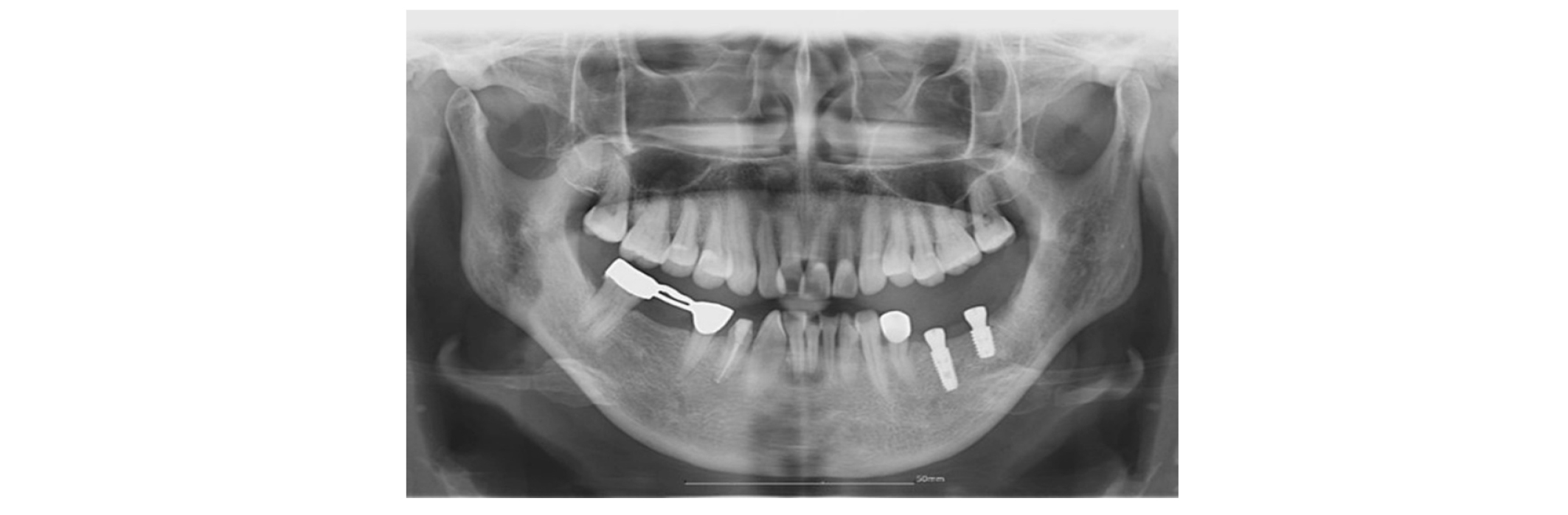
Fig. 3
Panoramic radiograph after implant installation.
Jae-Won Choi et al. : Implant-Supported Fixed Dental Prostheses with New Retention Type Using Zirconia Ball and Nickel-Titanium Spring.
Implantology 2019
임플란트 식립 3개월 째 임플란트의 안정성 변화를 추적 조사하기 위해 공진 주파수 측정 장치(Osstell Mentor, Integration Diagnostics AB, Göteborg, Sweden)를 이용하였으며, #35:83, #36:85 임플란트 안정성 지수(implant stability quotient; ISQ)값을 얻어 안정적인 골유착이 진행되었음을 확인하였다. 제조사의 지시에 따라 30 Ncm 조임회전력으로 abutment (Easy abutment, Samwon DMP, Yangsan, Korea)를 임플란트에 체결하였다. 인상용 코핑(Easy impression coping, Samwon DMP, Yangsan, Korea)을 abutment에 연결하고(Fig. 4), 실리콘 인상재(ImprintTM II GarantTM, 3M ESPE, Seefeld, Germany)와 개인 트레이를 이용하여 픽업 인상을 채득하고 주모형을 제작하였다(Fig. 5). 안궁이전 및 중심위를 채득하여 주모형을 교합기에 부착하였다(Fig. 6). 보철물 제작을 위해, lab analog (Easy lab analog, Samwon DMP, Yangsan, Korea)에 attachment (Easy abutment cylinder, Samwon DMP, Yangsan, Korea)를 연결하고 스캔 전 반사방지 파우더(Easy Scan, DMAX, Daegu, Korea)를 뿌린 후 모델 스캐너 (Identica Blue, Medit, Seoul, Korea)를 이용하여 디지털 인상을 채득하였다. 통상의 방법대로 computer aided design/computer aided manufacturing (CAD/CAM)을 이용하여 monolithic zirconia fixed dental prosthesis를 제작하였다(Fig. 7).
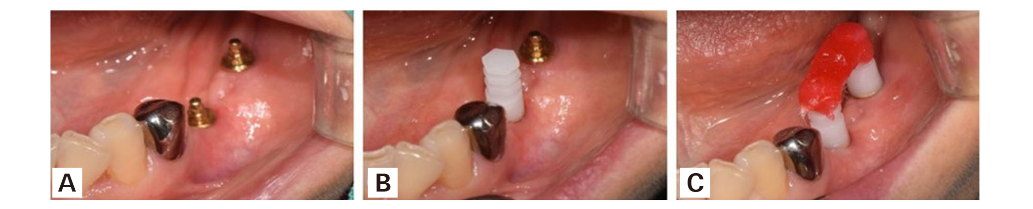
Fig. 4
(A) Abutment positioned onto implant, (B) Connecting impression coping to abutment, (C) splinting impression copings with pattern resin.
Jae-Won Choi et al. : Implant-Supported Fixed Dental Prostheses with New Retention Type Using Zirconia Ball and Nickel-Titanium Spring. Implantology 2019

Fig. 5
(A) Impression taking, (B) Fabrication of definitive cast.
Jae-Won Choi et al. : Implant-Supported Fixed Dental Prostheses with New Retention Type Using Zirconia Ball and Nickel-Titanium Spring. Implantology 2019
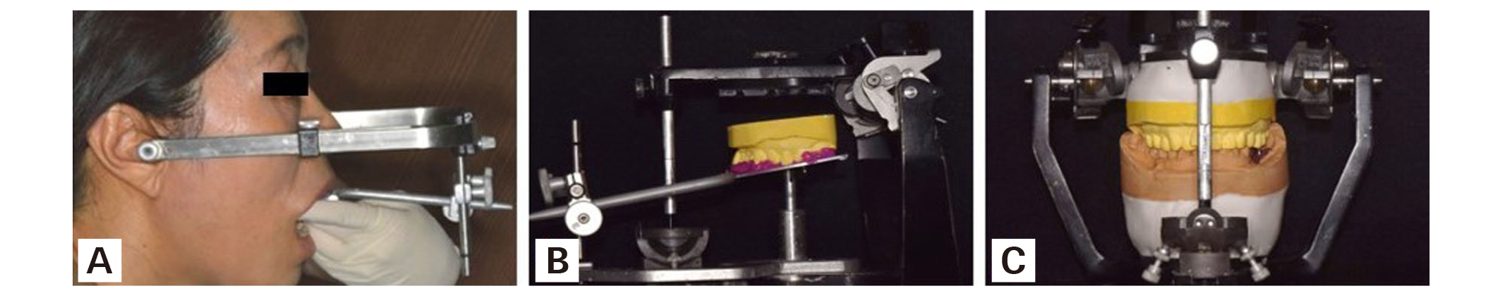
Fig. 6
(A) Facebow transfer, (B) Maxillary cast placed on the bite fork, (C) Mounting casts on the articulator.
Jae-Won Choi et al. : Implant-Supported Fixed Dental Prostheses with New Retention Type Using Zirconia Ball and Nickel-Titanium Spring. Implantology 2019
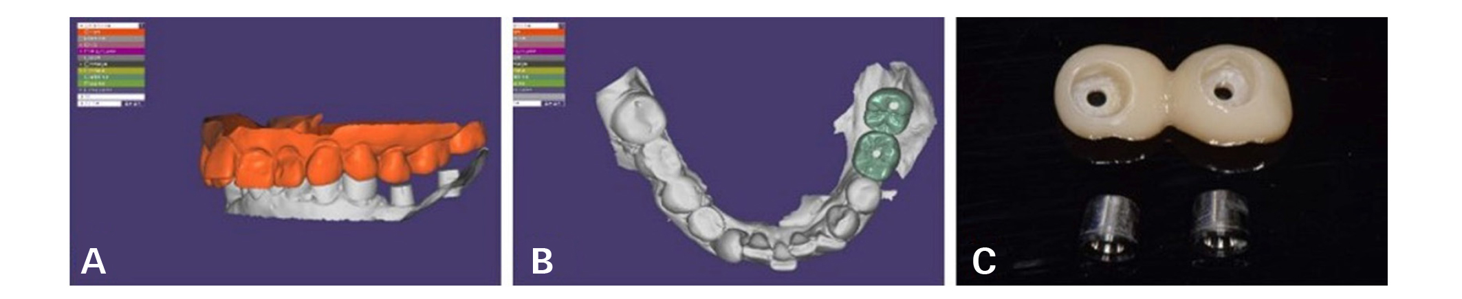
Fig. 7
(A) 3D model of the cast scanned using a 3D scanner, (B) Design of 2-unit zirconia bridge, (C) Definitive prosthesis.
Jae-Won Choi et al. : Implant-Supported Fixed Dental Prostheses with New Retention Type Using Zirconia Ball and Nickel-Titanium Spring. Implantology 2019
보철물을 최종 합착하기 전에 먼저 구강 내에 시적하여 적합, 심미성, 교합 등을 평가하였으며(Fig. 8), 자가 접착 레진 시멘트(G-CEM LinkAce, GC America, Alsip, IL, USA)를 이용하여 attachment에 최종 합착하였다(Fig. 9). 교합면 나사 구멍 주위의 잉여 시멘트를 제거하고 전용의 removal driver (Easy abutment remover driver, Samwon DMP, Yangsan, Korea)를 이용하여 보철물을 제거하였다(Fig. 10). 구강 외에서 보철물 치경부(cervical area) 주위의 잔류 시멘트를 제거하고 연마를 시행하였다. 보철물을 구강 내에 재시적하고 유동성 복합 레진(Filtek Z350 XT, 3M ESPE, MN, USA)으로 교합면 나사 구멍을 채워 최종 마무리하였다(Fig. 11).
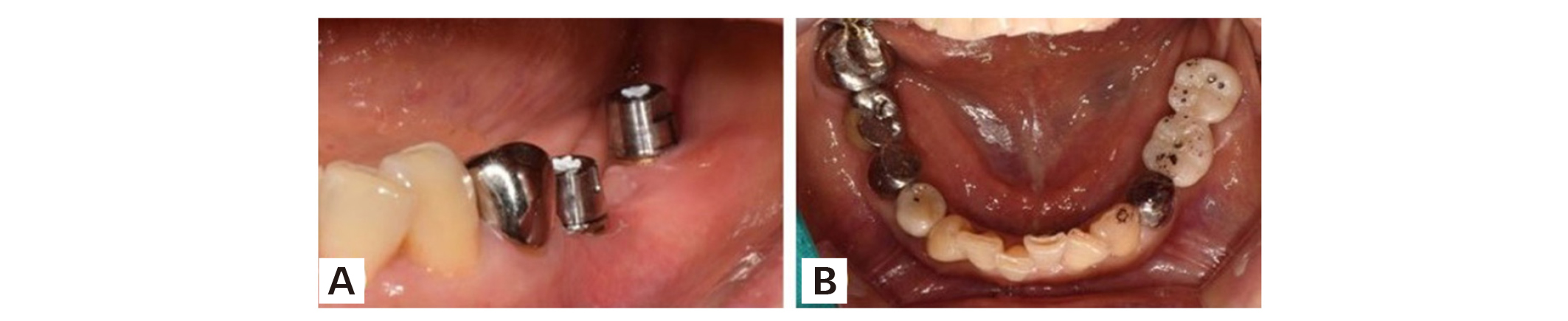
Fig. 8
(A) Attachment positioned onto abutment, (B) Inspection of prosthesis before final cementation.
Jae-Won Choi et al. : Implant-Supported Fixed Dental Prostheses with New Retention Type Using Zirconia Ball and Nickel-Titanium Spring. Implantology 2019
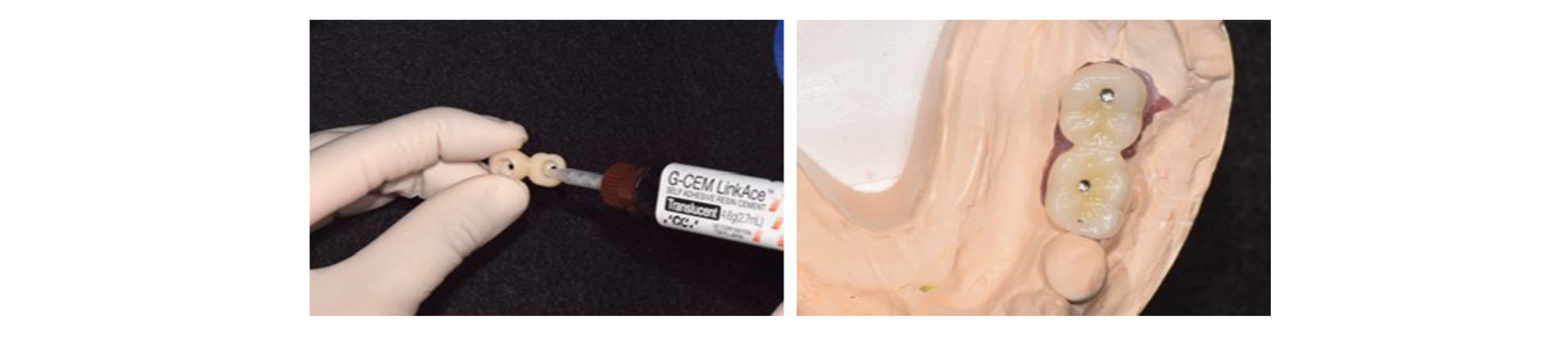
Fig. 9
Final cementation in definitive cast.
Jae-Won Choi et al. : Implant-Supported Fixed Dental Prostheses with New Retention Type Using Zirconia Ball and Nickel-Titanium Spring. Implantology 2019
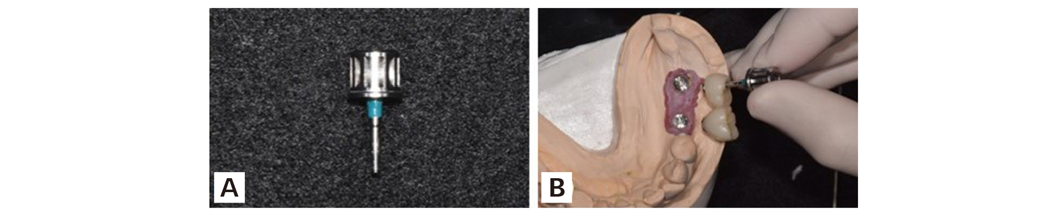
Fig. 10
(A) Removal driver, (B) Removal of prosthesis using removal driver.
Jae-Won Choi et al. : Implant-Supported Fixed Dental Prostheses with New Retention Type Using Zirconia Ball and Nickel-Titanium Spring. Implantology 2019
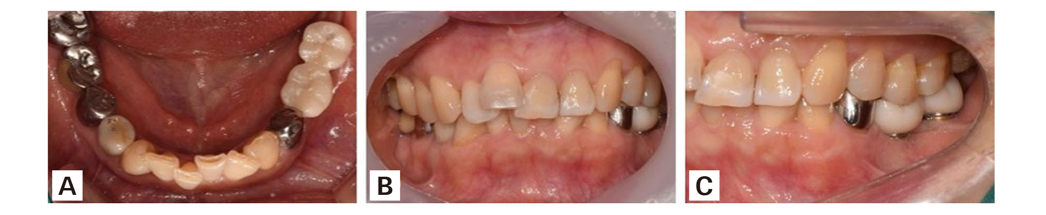
Fig. 11
Intraoral view after definitive prosthesis installation. (A) Occlusal view, (B) Frontal view, (C) Lateral view.
Jae-Won Choi et al. : Implant-Supported Fixed Dental Prostheses with New Retention Type Using Zirconia Ball and Nickel-Titanium Spring. Implantology 2019
Ⅲ. 총괄 및 고찰
본 증례에서는 새로운 유지 형태의 임플란트 보철 시스템인 EZC를 이용하여 zirconia fixed dental prosthesis를 제작하였다. EZC는 보철물을 retention screw없이 체결이 가능하고, 보철물의 최종 합착 후 구강 외에서 잉여 시멘트를 제거할 수 있기 때문에 기존의 임플란트 보철물 유지형태의 합병증이 발생하지 않는다는 장점이 있다. 또한, abutment의 헤드부는 편측으로 15° 테이퍼하기 때문에 임플란트가 평행하게 식립되지 못한 경우 임플란트 각도를 보상해줄 수 있다. 따라서 EZC는 단일치는 물론 본 증례와 같이 2개 이상의 임플란트를 이용한 고정성 보철물 수복 시에도 유용하게 사용할 수 있다.
EZC의 유지는 attachment의 지르코니아 볼이 abutment의 유지구의 언더컷(undercut) 부위에 위치하면서 가능해진다. 이 때, 지르코니아 볼의 외부에 있는 니켈-티타늄 스프링이 지르코니아 볼에 일정한 외력을 가함으로써 보철물은 안정적인 유지력을 보인다. 이는 니켈-티타늄 스프링이 넓은 작동 범위와 낮은 탄성 계수를 가지고, 스테인리스 스틸(stainless steel), 크롬-코발트(chromium-cobalt)와 같은 다른 합금보다 훨씬 더 탄력성을 띄는 martensitic-stabilized alloys로 가공되었기 때문이다17, 18. 또한, abutment와 attachment가 우수한 적합성을 가지도록 가공되어 있으며, 상부보철물이 이에 부착됨으로써 시멘트 유지형의 장점인 passive fit을 얻을 수 있게 한다. 이는 임플란트 식립 후 즉시 혹은 조기 부하 시 보철적 고려사항에서 장점이 될 수 있다. 볼의 주성분이 생체적합성, 굴곡 강도, 파절 강도, 내마모성과 같은 우수한 기계적 물성이 가진 지르코니아(ZrO2)라는 것도 이러한 안정적인 유지력에 영향을 끼칠 수 있다19. 또한 실제로 보철물을 구강 내 장착 이후 6개월간 추적관찰 동안 보철물의 탈락은 없었고 6개월 후에도 손의 힘으로는 쉽게 제거되지 않았다.
한편, retention screw에 의해 유지를 얻는 나사 유지형 보철물은 보철물을 제거하기 위해 교합면 나사 구멍이 필연적으로 형성되어야 하지만, 지르코니아 볼과 니켈-티타늄 스프링에 의해 유지를 얻는 EZC은 나사 구멍을 형성하지 않아도 보철물의 제거가 가능하다. 따라서 EZC는 보다 안정적인 교합 접촉과 더불어 심미적, 기계적으로 우수한 보철물을 제작할 수 있다. 다만, 본 증례와 같이 EZC에서도 술자의 선호도에 따라 교합면에 구멍을 형성할 수 있다. 이 구멍을 통해 전용의 removal driver를 attachment의 나사선에 따라 조이면, removal driver의 tip이 abutment의 상부를 밀게 됨으로써 최종적으로 보철물이 제거된다. 이처럼 전용의 removal driver를 사용하여 보철물을 제거할 경우 환자의 불편함을 최소화할 수 있고 다른 구성품에 무리없이 효율적으로 보철물을 제거할 수 있다. EZC의 경우, 교합면에 구멍을 형성한다고 할지라도 removal driver의 tip의 접근을 허용하기 위한 작은 직경(1.5 mm)만을 필요로 하기 때문에 상대적으로 큰 직경이 필요한 나사 유지형 보철물(3 mm)보다 교합 및 심미성에 있어서 유리하다.
6개월의 추적관찰 후, 정상적인 구강 기능하에서 EZC로 제작된 보철물의 탈락이나 파절 양상은 보이지 않았으며, 심미적 및 기능적 측면에서의 환자 만족도 역시 전반적으로 우수하였다. 임플란트 주위조직의 건강상태는 양호하였으며, 염증, 부종, 궤양과 같은 증상 또한 발견되지 않았다. 하지만, abutment와 보철물 사이에 음식물이 저류된 흔적(food collection)의 문제들이 관찰되기도 하였다. 아직까지 EZC는 내부 연결형 임플란트 고정체에만 한정적으로 적용 가능하고 attachment의 구조상 맞춤형으로 제작할 수 없으며, 전용의 기구가 필요하다는 한계가 있다. 따라서 EZC의 문제점과 한계를 극복할 수 있는 수정 및 개선의 노력과 더불어 장기적 관점에서의 추가적인 임상 연구가 이루어져야 할 것이다.
Ⅳ. 결론
EZC는 지르코니아 볼과 니켈-티타늄 스프링이 보철물의 유지를 담당하므로 retention screw가 필요 없어 screw와 관련된 기계적 문제가 없고 착탈이 간편하며, 지속적인 유지력을 제공한다. 또한, 보철물의 최종 합착 후 시멘트를 구강 외에서 제거할 수 있어 잔류 시멘트로 인한 생물학적 합병증을 방지할 수 있다. 다양한 이점이 있는 새로운 유지 형태의 EZC는 임플란트 보철물 수복에 있어서 향후 활용 가능성이 높을 것으로 기대되며, 추후 장기적인 임상 시험을 통해 임상적 유용성을 검증해야 할 것이다.
Acknowledgements
본 연구는 보건복지부의 재원으로 한국보건산업진흥원의 보건의료기술 연구개발사업 지원에 의하여 이루어진 것임(과제고유번호 : HI18C0594).
References
Levine RA, Clem D, Beagle J, et al. Multicenter retrospective analysis of the solid-screw ITI implant for posterior single-tooth replacements. Int J Oral Maxillofac Implants. 2002; 17: 550-556.
Hebel KS, Gajjar RC. Cement-retained versus screw-retained implant restorations: Achieving optimal occlusion and esthetics in implant dentistry. J Prosthet Dent. 1997; 77: 28-35.
Lee A, Okayasu K, Wang HL. Screw- versus cement-retained implant restorations: Current concepts. Implant Dent. 2010; 19: 8-15.
Shadid R, Sadaqa N. A comparison between screw- and cement-retained implant prostheses. A literature review. J Oral Implantol. 2012; 38: 298-307.
Ma S, Fenton A. Screw- versus cement-retained implant prostheses: A systematic review of prosthodontic maintenance and complications. Int J Prosthodont. 2015; 28: 127-145.
da Rocha PV, Freitas MA, de Morais Alves da Cunha T. Influence of screw access on the retention of cement-retained implant prostheses. J Prosthet Dent. 2013; 109: 264-268.
Pauletto N, Lahiffe BJ, Walton JN. Complications associated with excess cement around crowns on osseointegrated implants: a clinical report. Int J Oral Maxillofac Implants. 1999; 14: 865-868.
Weber HP, Kim DM, Ng MW, et al. Peri-implant soft-tissue health surrounding cement- and screw-retained implant restorations: A multi-center, 3-year prospective study. Clin Oral Implants Res. 2006; 17: 375-379.
Zarone F, Sorrentino R, Traini T, et al. Fracture resistance of implant-supported screw- versus cement-retained porcelain fused to metal single crowns: SEM fractographic analysis. Dent Mater. 2007; 23: 296-301.
Heo YK, Lim YJ. A newly designed screw- and cement-retained prosthesis and its abutments. Int J Prosthodont. 2015; 28: 612-614.
Chung CH, Son MK. The classification and comparison of implant prosthesis according to types of retention. Part I: Screw retained prosthesis vs cement retained prosthesis. Implantology. 2010; 14: 138-151.
Taylor TD, Agar JR. Twenty years of progress in implant prosthodontics. J Prosthet Dent. 2002; 88: 89-95.
Chee W, Felton DA, Johnson PF, et al. Cemented versus screw-retained implant prostheses: Which is better? Int J Oral Maxillofac Implants. 1999; 14: 137-141.
Michalakis KX, Hirayama H, Garefis PD. Cement-retained versus screw-retained implant restorations: A critical review. Int J Oral Maxillofac Implants. 2003; 18: 719-728.
Yoon NR, Lee SB, Lee SW, et al. A new retaining method of cement-retained restoration with linguo-horizontal insertion of fiber post. J Korean Acad Prosthodont. 2017; 55: 71-78.
Choi JW, Choi KH, Chae HJ, et al. Load-bearing capacity and retention of newly developed micro-locking implant prosthetic system: An in vitro pilot study. materials (Basel). 2018; 11: 564.
Ferreira MA, Luersen MA, Borges PC. Nickel-titanium alloys: A systematic review. Dental Press J Orthod. 2012; 17: 71-82.
Gurgel Jde A, Kerr S, Powers JM, et al. Torsional properties of commercial nickel-titanium wires during activation and deactivation. Am J Orthod Dentofacial Orthop. 2001; 120: 76-79.
Manicone PF, Rossi Iommetti P, Raffaelli L. An overview of zirconia ceramics: Basic properties and clinical applications. J Dent. 2007; 35: 819-826.


